, James B. Galloway2 and David L. Scott2
(1)
Molecular and Cellular Biology of Inflammation, King’s College London, London, UK
(2)
Rheumatology, King’s College Hospital, London, UK
Abstract
The hallmark of the inflammatory arthropathies is joint inflammation, characterised by joint pain and swelling. Different disease types affect different joints. RA classically affects the peripheral small joints in a symmetrical fashion. By contrast the seronegative spondyloarthropathies tend to affect the spine and peripheral large joints in an asymmetrical fashion. Extra-articular features are common in all forms of inflammatory arthritis. In RA almost any body system can be affected, with manifestations ranging from rheumatoid nodules to mononeuritis multiplex. The classical extra-articular manifestation of the seronegative spondyloarthropathies is anterior uveitis. This chapter will discuss the clinical manifestations of the inflammatory arthropathies, spanning their articular and extra-articular features.
Keywords
SynovitisTenosynovitisTender JointsEnthesitisExtra-Articular InvolvementSynovitis of Peripheral Joints
Symptoms
The main symptoms of inflammatory arthritis result from inflammation of the joints. Pain is a dominant symptom both within the joints and more diffusely around the joints. The joints are also swollen and tender and they are difficult to move.
The symptoms often show diurnal variation. Patients have most problems early in the morning. As a result they usually have prolonged morning stiffness. This can last up to several hours. It usually lasts over 30 minutes. The exact nature of the stiffness can be difficult to define and not all patients describe it in the same manner.
Joint Swelling and Tenderness
The characteristic features of inflamed joints are swelling and tenderness [1]. Joint swelling is soft tissue swelling detected along joint margins. When there is a synovial effusion, a joint is inevitably swollen. However, effusions are not mandatory features of a swollen joint. The characteristic feature of a swollen joint is fluctuation, when fluid is displaced by pressure in two planes.
Bony swelling and joint deformities often complicate the counting of swollen joints. Neither of these indicates the presence of joint swelling, although they can be present if joints are swollen. In late disease it is often difficult to differentiate swollen from deformed inactive joints.
An associated clinical feature that accompanies joint swelling is swelling of tendon sheaths – termed tenosynovitis. It involves an identical pathological process.
Joint tenderness is indicated by inducing pain in a joint at rest with pressure. Judging the correct amount of pressure to elicit tenderness depends on both the examiner and the patient. Generally sufficient pressure should be exerted by the examiner’s thumb and index finger to cause ‘whitening’ of the examiner’s nail bed. Too little pressure may fail to elicit joint tenderness even though it is present. Too much pressure will result in pain in everyone. The level of pressure required to elicit joint tenderness varies from one patient to another. In some joints, like the hip, tenderness is best identified through movement.
Peripheral Joints Involved
RA is usually a symmetrical polyarthritis. It mainly involves the hand, wrists, feet, and some large joints, particularly the knees. In some patients it only involves a few joints at the outset, though more become involved over time [2, 3].
Other forms of inflammatory arthritis usually involve fewer joints. The distribution of joint involvement is also less obviously symmetrical [4]. Each form of inflammatory arthritis has its own relatively characteristic distribution of joint involvement.
Rheumatoid Arthritis
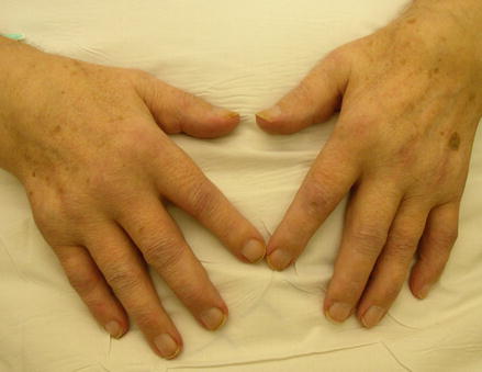
Hand involvement is particularly characteristic of RA (Fig. 3.1). It usually involves the metacarpophalangeal, proximal interphalangeal, thumb interphalangeal and wrist joints. The distal interphalangeal joints can be affected when there is coexisting disease in other hand joints.


Figure 3.1
An inflamed hand in early rheumatoid arthritis
Involvement of the tendons is also characteristic. Tenosynovitis of flexor tendons reduces finger flexion and strength. Nodular thickenings in tendon sheaths may result in a trigger finger.
Damage to the wrists causes compaction of bone at the small wrist joints. In late disease this damage may progress to bony ankylosis. Historically in late RA characteristic deformities developed in the hands. There was ulnar deviation of the fingers, subluxation of the metacarpophalangeal joints, hyperextension of the proximal interphalangeal with flexion of the distal joints (swan-neck deformity), flexion of the proximal interphalangeal with hyperextension of the distal joints (boutonnière deformity), and a Z-shaped deformity of the thumb. These changes are seen less often in the modern era.
The small joints of the feet are involved at an early stage, causing considerable difficulty walking. As the disease progresses a complex series of changes occurs in the feet including spreading of the forefoot, dorsal subluxation of toes and subluxation of metatarsal heads to a subcutaneous site on the plantar surface. In some cases additional hallux valgus leads to “stacking” of the second and third toes on top of the great toe.
The ankle joint itself is rarely involved in RA, although it is sometimes damaged in late disease. By contrast the subtalar joint is often involved. Its involvement results in pronation deformities and eversion of the foot. The reason for such a different level of involvement in adjacent joint is most likely the effects of microtrauma on the severity of synovitis. The ankle is in a relatively atraumatic environment compared to the subtalar joint.
Knee involvement is common in RA including both early and late disease. Quadriceps wasting and loss of full extension both occur in the first stages of the disease.
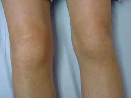
In some patients there may be large effusions (Fig. 3.2), and these can result in a popliteal or Baker’s cyst. Synovial fluid entering these popliteal cysts does not readily return into the knee. One consequence is that high pressures may be generated and the cyst can rupture into the calf, resulting in considerable pain and discomfort.


Figure 3.2
Knee involvement in rheumatoid arthritis
RA often affects the synovium of the glenohumeral joint in the shoulder and its associated bursae. In addition it involves the rotator cuff together with its associated muscles on the chest wall. Weakness of the rotator cuff apparatus may result in shoulder subluxation.
The hips are rarely involved in the early stages of RA. However, their involvement is more frequent in late disease. Eventually about half of rheumatoid patients develop some evidence of hip disease. About 20 % of patients develop significant levels of hip pain with resulting joint failure.
In a small number of patients the femoral head collapses. The acetabulum is remodelled and pushed medially. This results in protrusio acetabuli. The deformity usually progresses until the femoral neck impinges on side of pelvis.
Cervical pain is common in the early phases of RA. This pain is mainly due to muscle spasm. However, over the subsequent course of their disease up to 90 % of patients develop some cervical spine involvement. The likelihood of severe cervical spine involvement is increased in long standing disease and when many joints are involved. Significant subluxations occur in about one third of cases. Neurological deterioration can be irreversible in such cases, and it is therefore important to look for subtle signs of early neurological involvement. In addition to painful limitation of neck motion, warning signs of significant cervical involvement include sub-occipital pain, paraesthesia in hands and feet, urinary retention and incontinence and involuntary leg spasms.
Psoriatic Arthritis
PsA has highly variable clinical features and there are several different patterns of joint involvement [5]. These tend to change over time.
Conventionally PsA is divided into five different clinical subtypes. These clinical subtypes comprise:
1.
Polyarthritis: many joints are involved and the clinical pattern is very similar to RA
2.
Oligoarthritis: in most patients with PsA only two or three joints are involved. As a consequence the arthritis seems very asymmetric.
3.
Monoarthritis: as the classification indicates only one joint is involved. Over time the pattern may change with the involvement of more joints.
4.
Distal interphalangeal joint arthritis: this is a highly characteristic arthritis of the distal interphalangeal joints of the hands. It can be very destructive.
5.
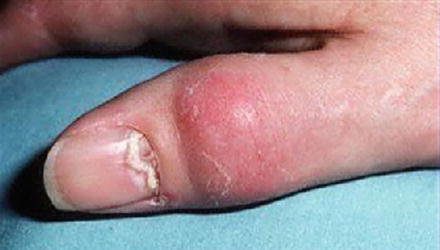
Arthritis mutilans: this is the rare but highly progressive and destructive form of arthritis which is seen in occasional patients (Fig. 3.3).


Figure 3.3
Psoriatic arthritis involving the thumb joint
Other Forms of Spondyloarthropathies
Peripheral joint inflammation can be the presenting feature in many patients with a spondyloarthritis. It can also occur against the background of established inflammatory back disease in AS.
In these seronegative types of arthritis usually only one or two joints are involved. Patients have an oligoarthritis, which is mainly seen in lower limbs. It is usually asymmetric in distribution. The most commonly involved joints are the knees. In some patients the elbows can also be involved.
Enthesitis
The entheses are the insertion of tendons into bone [6]. These are frequently inflamed in spondyloarthritis. Such inflammation is known as an enthesitis. It involves sites such as the insertion of the Achilles tendon – Achilles tendonitis – and insertion of the plantar fascia – plantar fasciitis. Other sites involved include the greater trochanters, the pelvic brim, and the epicondyles.
Stay updated, free articles. Join our Telegram channel

Full access? Get Clinical Tree


