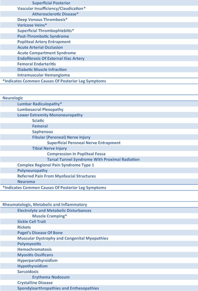
Fig. 6.1
Differential diagnosis of posterior leg pain, weakness, or swelling
The goal of this chapter is to provide a functional and practical guide to the physical examination of the posterior leg. The level and scope of the material presented is intended to be inclusive of the needs of a variety of health-care professionals. In general, physicians need to have a broader scope with an eye towards referred pain or signs of systemic medical problems while rehabilitation professionals require a more detailed working knowledge of musculoskeletal etiologies for pain and dysfunction. The chapter is organized into sections covering the standard examination areas: inspection, gait and station, palpation, range of motion (ROM), strength testing, neurologic status, vascular status, and special tests. An attempt has been made to correlate exam finding with the associated diagnosis in the differential.
Acute injuries often necessitate a more circumscribed examination due to pain and more limited goals of determining the specifics of the injury location and severity. In acute injuries, it is also important to evaluate for potential complications and be cognizant of confounding or coexisting injuries. For chronic or sub-acute complaints, a more comprehensive and functional approach is required. Particular attention should be paid to predisposing factors for injury and referred pain from neurologic, vascular, and bony pathology. There is some wisdom in the playful joke that the only aptitude needed to do orthopedics is the capability to compare one side of the body to the other. So whenever possible, take advantage of the natural symmetry of the body to compare apparent abnormal finding relative to the contralateral limb.
Inspection
It is important to completely visualize the bilateral lower extremities during the examination. This is best accomplished by having the patient disrobe or change into examination shorts prior to beginning the evaluation. During inspection careful attention should be paid to signs of bruising, swelling, and skin changes that may suggest underlying vascular or neurologic disease. Visible erythema and swelling associated with pain and increased warmth are the classic signs of inflammation and should prompt the clinician to consider inflammatory causes of leg pain. Inflammatory causes of leg pain can be localized or systemic. Leg pain in the presence of skin rashes and nail abnormalities are also suggestive of possible systemic inflammatory conditions. Scars or wounds on the legs should be noted and correlated with the past medical and surgical history.
If bruising is found, note the location, size, distribution, and color. Location and distribution of ecchymosis can suggest the location of the injury and its depth. The color of the bruising suggests the acuity of the bleeding. Upon extravasation of blood, hemoglobin begins to degrade in a sequential fashion, giving rise to progressive change in color and eventual resolution of bruising by 2 weeks. In this way, the color of the bruise can be used to age the injury: red-blue color (0–5 days), green color (5–7 days), yellow color (7–10 days), and golden-brown color (10–14 days). It should be noted that bruising or contusion refers to subcutaneous bleeding that has a traumatic etiology. Ecchymosis is a more general description of subcutaneous bleeding greater than 1 cm and may not be traumatic in etiology. Nontraumatic causes of ecchymosis include coagulopathies. Subcutaneous bleeding less than 1 cm in diameter is either called petechia (< 3 mm) or purpura (3 mm–1 cm), which are associated with platelet disorders or vasculitis, respectively. Bruising is easier to identify in patients with light skin complexions. It is common for the blood to travel along soft tissue planes which often results in migration of the bruising over time.
If calf swelling is present, take note if the calf swelling is unilateral or bilateral. Unilateral calf swelling is indicative of localized pathology whereas symmetrical swelling suggests a systemic cause. Inspection can also help determine if the swelling can be classified as pitting or non-pitting edema. Indentations from socks, shoes, or other tight-fitting clothing can be useful in detecting pitting edema. Non-pitting edema is caused by lymphedema. Pitting edema has a variety of local and systemic causes. Acute unilateral pitting edema of the leg is often due to deep venous thrombosis (DVT), cellulitis, a ruptured bakers cyst, or trauma.
Stigmata of chronic vascular disease should also be sought on inspection . Arterial peripheral vascular disease should be suspected if there are well-demarcated ulcers or non-healing lesions in the distal extremity along the ground contact areas, erythema of the foot in a dependent position which changes to pallor with elevation, and skin that is shiny, dry, and atrophic with absence of hair. Chronic venous disease is suspected on inspection if chronic soft, pitting edema is seen in the ankle and evidence of dependent varicosities of the leg with hyperpigmented skin and venous ulcerations over medial malleolus.
Inspection of the legs may also reveal stigmata of neuropathy. Thin ankles and legs along with deformities of the arch and toes can suggest hereditary neuropathies such as Charcot–Marie–Tooth (CMT) disease. Complex regional pain syndrome type 1 is associated with visible swelling and discoloration of the affected leg.
Screening for leg length should be done on inspection. A reasonable screening test of leg length symmetry can be done by observing the patient from behind while comparing the relative height of popliteal and gluteal creases. If you suspect a clinical relevant difference in leg length, additional clinical tests should be performed for further assessment. Specific examination techniques for leg length are discussed below.
Observation of the lower extremities during functional movement is important. At a minimum, inspection should be done on standing and walking. In addition, observation while running or other relevant movements can greatly aid the clinical evaluation. Evaluation of gait and station are covered separately in the next section.
Evaluation of Gait and Station
Examination of gait and station should be incorporated with the examination of the lower extremity. Because of the functional role the muscles of the posterior leg have in standing and ambulating, injuries to these muscles are often most noticeable during the evaluation of gait and station. Correlating abnormalities in gait and station to the underlying pathology requires an understanding of the normal and abnormal presentation. It should be noted that abnormities of gait and station not only can be the result of pathology but also can be causative factor in precipitating the injury. Therefore, although evaluation of gait and station can help the clinician in the diagnosis of acute muscle injuries in the posterior leg , the detailed evaluation of gait is most helpful in investigating chronic or recurrent issues that may be caused by underlying biomechanical abnormalities.
Station or stance position is evaluated by observation of the standing patient. Normal station should reveal a balanced body posture over the center of gravity and symmetrical alignment of the lower limbs and feet. In neutral stance position, alignment of the lower limb should reveal a weight-bearing line in a sagittal plane through the anterior superior iliac spine, patella, and second metatarsal of each lower extremity. The heel should be perpendicular to the forefoot and in line with the tibia. The knee should be fully extended with slight valgus alignment. The subtalar joint and hips should be in a neutral position. The pelvis should exhibit a slight anterior tilt. Significant deviations from ideal stance position suggest structural abnormalities or compensations that may lead the clinician to the diagnosis or suggest the cause of the presenting complaint (Table 6.1) . When these common clinical associations are subject to evaluation by evidence-based methods the conclusions reached have been very limited [2, 3].
Table 6.1
Biomechanical abnormalities commonly associated with posterior leg injury
Biomechanical abnormality | Posterior leg injury |
|---|---|
Excessive pronation (dynamic pes planus) | Achilles tendinopathy |
Medial tibial stress syndrome | |
Tibial stress fracture | |
Fibular stress facture | |
Tibialis posterior injury | |
Flexor hallucis longus injury | |
Excessive supination (pes cavus) | Tibia stress fracture |
Fibular stress fracture | |
Hindfoot varus | Medial shin pain |
Fibular stress facture | |
Ankle equinus (restricted ankle dorsiflexion) | Achilles tendinopathy |
Medial tibial stress syndrome | |
Calf strain | |
Stress fractures |
Gait is classically described by its two component actions called the stance phase and the swing phase. The stance phase refers to the portion of gait when the foot is on the ground, while the swing phase refers to the portion of gait when the foot is no longer in contact with the ground. The repeated progression of stance phase through swing phase produces the motion of walking and running called the gait cycle. The phases of gait are further divided into component actions. The stance phase is subdivided into heel strike, mid-stance, and push-off. The swing phase is subdivided into acceleration, mid-swing, and deceleration.
The phases of running and walking are similar. Running is defined by the addition of a flight phase between stance phases in which neither foot is in contact with the ground. While walking, 60 % of the gait cycle is spent in stance phase and 40 % in swing phase. During running, the portion of time spent in stance phase decreases with increasing running speed. In addition, running is associated with greater ROM of the pelvis, hip, and knee as well as a narrower base of gait when compared to walking.
Examination of gait can begin even before the formal exam. Observation of the patient as they ambulate through the office is most likely to reveal natural movement patterns and can quickly identify an obvious limp or the use of assistive devices. During formal examination of gait, try to identify which phase of gait and component motion is pathologic. Most abnormalities will be present in stance phase where the extremity is susceptible to the liabilities of weight bearing. Patients with muscle weakness in the gastrocnemius , soleus, and flexor hallucis longus or Achilles rupture may exhibit a flat foot gait with no or limited push-off. Gastrocnemius strains may result in the patient adopting a gait characterized by excessive knee flexion and ankle plantar flexion in an attempt to splint the affected muscle. A bouncy gait suggests early heel lift from ankle equinus or heel pain. Compensations related to ankle equinus lead to posterior leg muscles strains, Achilles tendon injuries, shin splints, and stress fractures. Patients with fractures or other severe acute trauma will have pain in all phases of gait and are likely to either not bear weight on the affected extremity or spend as little time as possible in stance phase.
During mid-stance, as the body passes over the weight-bearing limb, the pelvis and trunk shift laterally over the weight-bearing side and drop slightly on the non-weight-bearing limb. Excessive lateral displacement during mid-stance is caused by gluteus medius weakness and results in a gluteus medius or abductor lurch gait. The Trendelenburg test evaluates the function of the gluteus medius in maintaining dynamic pelvic stability in single-leg, weight-bearing stance. The test is performed by observing the patient from behind while they are standing in double-leg stance and then in single-leg stance alternating between each leg. A positive test occurs when the pelvis does not elevate on the unsupported side during single-leg, weight bearing stance. A positive test indicates inadequate force is being generated by the gluteus medius on the stance leg to maintain dynamic pelvic stability during single-leg, weight bearing stance. This can lead to biomechanical abnormalities which increase likelihood of injuries. Inadequate gluteus medius force generation can be caused by poor gluteus medius development, poor activation sequence, nerve root mediated weakness, and anatomical variations that functionally shorten the muscle.
Excessive subtalar pronation can lead to overload of the soleus , gastrocnemius, and tibialis posterior during dynamic supination and plantar flexion. This can lead to excessive muscle tightness and soreness. In addition, overload from excessive subtalar pronation can predispose the muscles of the posterior leg to strain and tendon damage. Moreover, excessive muscle overload can cause muscular hypertrophy that can contribute to the development of compartment syndrome.
Excessive supination of the subtalar joint suggests contracture of the gastrocnemius, soleus, and tibialis posterior, structural abnormalities of the foot or weak peroneals. Excessive supination limits mobility of the foot resulting in reduced ability to attenuate force during the gait cycle and increased risk for stress fractures of the leg and foot.
Palpation
Bony Palpation
Palpation of the bones of the posterior leg is limited due to the predominance of soft tissue structures. However, palpation of bones remains an important part of the clinical examination of the posterior leg, and palpation of relevant bony landmarks of the knee and ankle is also significant. Examination by palpation is best performed with the patient seated on the edge of the examination table. This reduces the muscular tone of the leg muscles and allows the knee and ankle to be manipulated for optimal visualization and palpation of the bony landmarks. If the patient cannot sit, then it is best to position the patient supine with the knee flexed to 90°.
On the medial aspect of the leg, the tibia should be palpated along its length. Begin at the medial joint line to assess for medial joint line tenderness which can suggest osteoarthritis, medical meniscus pathology, and medial collateral ligament (MCL) strain or bursitis. Follow the medial tibia plateau distally to the distal medial collateral ligament and pes anserine insertion, then down along the medial tibia border to the medial malleolus. Focal pain along the medial tibial border can be indicative of stress fracture or if more diffuse periostitis. Chronic periostitis is often associated with palpable firm bumpy uneven texture along the midportion of the tibia. The posterior medial border of the tibia is palpable by digging the fingers around behind the medial border, carefully probing the posterior aspect of the tibia can help to identify stress fractures of the posterior cortex which can present as calf pain with or without shin pain. After palpating the distal aspect of the posterior tibia, examination should extend into the foot to probe the navicular tubercle. The navicular tubercle is the insertion of the posterior tibialis, and pathology of the navicular can often give rise to medial ankle and leg pain.
Bony palpation of the posterior leg is limited to the flabella of the lateral gastrocnemius tendon and posterior ankle. In the ankle, the posterior talus and dome of the calcaneus should be examined. The dome of the calcaneus and posterior talus are palpated in the soft depression just anterior to the Achilles tendon. Pain with bony palpation of the calcaneus can suggest fracture, stress injury, or in adolescents—calcaneal apophysitis. Compression of the calcaneus for each side, the so-called squeeze test, can help evaluate for bony pain. The pain with palpation of the posterior talus suggests possible posterior malleolus fracture, os trigonum syndrome, or posterior impingement syndrome . Deformities of the calcaneus and soft tissue calcification should be noted and palpated for tenderness. A Haglund’s deformity presents as a distinct bony prominence of the posterolateral calcaneus. This should be distinguished from Haglund’s disease which results from soft tissue inflammation of the retrocalcaneal bursa and Achilles tendon insertion. Enthesophytes or spurring at the insertion of the calcaneus is common and may be associated with pain on palpation.
Bony palpation of the lateral aspect of the leg should begin at the lateral joint line extending along the lateral tibial plateau to head of the fibula. Instability, arthritis, or ganglions of superior tibiofibular joint often present as pain in the lateral calf or leg. Continuing distally an attempt should be made to palpate the entire length of the fibula to the lateral malleolus. Focal pain with palpation of the fibula without a history of trauma suggests stress fracture, injuries that often refer pain to the posterior or lateral leg. The proximal fibula can also be fractured from a direct blow or following significant rotation ankle sprains that result in propagation of a Maisonneuve fracture .
Soft Tissue Palpation
Soft tissue palpation is often the most valuable part of the clinical examination for posterior leg pain. When you are familiar with the natural history of muscle injuries in the posterior leg, hear the patients’ account of their pain and observe them walk into the exam room; anatomical-based palpation can often quickly allow the examiner to literal and figuratively put their finger on the problem. Although this is very rewarding, it is critical to complete the exam to evaluate for any concomitant pathology and assess for causative factors.
It is important to palpate the soft tissue structures in their entirety. In respect to muscles, the whole muscle belly as well as the associated tendons and aponeurosis should be carefully examined for trigger points, increased tone, swelling, thickening, defect, or mass. It is helpful to palpate in both a relaxed state and during active contraction. Palpating though the full range of relative joint motion can aid evaluation of symptomatic muscles. In addition, some soft tissue palpation is best done after exertion. This is particularly helpful to evaluate for muscle herniations and chronic compartment syndrome where the palpation of the can be normal at rest but become tight, tender, and swollen with extended exertion.
To perform a comprehensive examination of the leg by palpation, it is helpful to proceed in a sequential fashion. Begin with the posterior aspect of the knee. Here the popliteal fossa can be a good landmark to help define the relevant structures. The superior borders of the popliteal fossa are bound by the biceps femoris tendon laterally and the semimembranous and semitendinosus muscle medially. From this location, distal biceps femoris should be palpated to its attachment on the posterior fibula and the medial hamstring tendons along the posterior medial knee to their respective attachments. The semitendinosus tendon travels medially with the sartorius and gracilis to form the combined pes anserine insertion. The location of the pes anserine is a common site of bursitis on the anterior medial leg but which can extend posteriorly causing leg pain and swelling.
The inferior margins of the fossa are formed by the medial and lateral heads of the gastrocnemius . When present, the plantaris muscle belly will be palpable medial to the lateral head of the gastrocnemius in the popliteal fossa. A neurovascular bundle containing the popliteal artery, vein, and posterior tibial nerve cross through the popliteal area from deep to superficial, respectively.
With the knee in a relaxed and flexed position, the popliteal artery pulse can usually be felt by pressing the fingers deep into the fossa along the posterior joint capsule of the knee. Absence or asymmetrical palpation of the popliteal pulse can suggest vascular occlusive disease or mechanical obstruction. Because it is the most superficial structure crossing the popliteal area, occasionally disorders of the posterior tibial nerve can result in a palpable neuroma or positive compression test for neuritis. The tibial nerve provides muscle and sensory innervation to the posterior leg. Isolated tibial neuropathy is uncommon but can arise from a schwannoma or direct compression from a popliteal mass such as a baker’s cyst, popliteal aneurysm, or hematoma .
The saphenous nerve supplies sensation to the medial leg. Saphenous nerve injury can be an isolated finding or related to femoral neuropathy. An isolated injury to the saphenous nerve will produce only sensory deficits. The saphenous nerve can be injured during knee surgery, venous grafting for coronary artery bypass, or from direct trauma. Injuries to the saphenous nerve can lead to tenderness or paresthesias in the medial leg. The infrapatella branch of the saphenous nerve can be palpated over the medial tibia at the level of the tibial tubercle. A positive Tinel’s sign can help confirm the diagnosis by briskly tapping along the nerve to reproduce symptoms.
Related posts:

Stay updated, free articles. Join our Telegram channel

Full access? Get Clinical Tree


