Fig. 7.1
Human arm kinematic relationships
The underlying kinematics are shown in Fig. 7.1 and include the wrist action considered in Chap. 6. The rehabilitation system is shown in Fig. 7.2 and facilitates recovery of upper limb motor control and function through goal-oriented, functional tasks assisted by ES. The stroke participant sits at the workstation and ES electrodes are positioned on the anterior deltoid, triceps and wrist and finger extensors of their impaired arm. If required, mechanical support is provided by the SaeboMAS unweighting device described in Sect. 2.1.3 that facilitates movement by supporting the arm against gravity. A Microsoft Kinect (Microsoft, Washington, USA) is used to provide shoulder, elbow and wrist joint positions, and has an accuracy of approximately 10 mm [3, 4]. It is combined with an electro-goniometer (Model SG75, Biometrics Ltd, Newport, UK) placed over the wrist joint to measure flexion/extension and abduction/adduction. Using these data, the joint angles shown in Fig. 7.1 are computed and used by the ES control scheme. A custom made graphical user interface is used to select appropriate tasks and monitor training. For safety purposes an over-ride ‘stop’ button terminated trials with immediate effect.
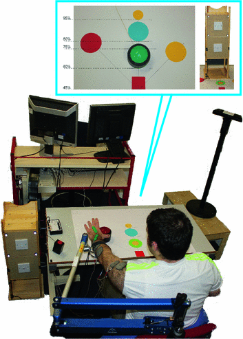
Fig. 7.2
Rehabilitation system incorporating SaeboMAS, Microsoft Kinect, task display, operator monitor and real-time control hardware. The bubble displays the task template customized to each participants arm length. Green  button located at 60 % of arm length; Blue
  button located at 80 % of arm length; Red
  button located at 75 % of arm length, 45
 to the impaired side; Yellow
  button located at 75 % of arm length, 45
 across body; small yellow circles
  location that object was grasped from and repositioned to (60 and 95 % of arm length). The cabinet housed the light switch tasks (located at 75 and 80 % of reach for the high and low light switch tasks respectively); the draw task (located at 80 % of reach) was on the reverse side of the cabinet
button located at 60 % of arm length; Blue button located at 80 % of arm length; Red button located at 75 % of arm length, 45 to the impaired side; Yellow button located at 75 % of arm length, 45 across body; small yellow circles location that object was grasped from and repositioned to (60 and 95 % of arm length). The cabinet housed the light switch tasks (located at 75 and 80 % of reach for the high and low light switch tasks respectively); the draw task (located at 80 % of reach) was on the reverse side of the cabinetThe rehabilitation system incorporates five main functional tasks that span a 3-dimensional workspace and offers a range of reaching and grasping challenge. They comprise closing a drawer, pressing a light switch (located at 90 or 115
 of shoulder elevation), stabilizing an object, pressing a button (placed at one of four different locations in the workspace) and lifting to reposition an object. Objects can be placed at different locations on the table corresponding to percentages of arm reach (60, 75, 80, 95 %), and either directly in line with the shoulder or 45
 to either side (see Fig. 7.2). The table was at a distance of 45 % of arm length away from the gleno-humeral joint and 35 cm below the arm when held 90
 horizontal to the shoulder. These values were used to define the extended task tracking problem (6.4) of Chap. 6 which is then solved by following control design Procedure 4. Since only the reaching component was used, the button pressing and drawer closing tasks were defined as in Sect. 6.6 with parameter T modified for each participant. Repositioning of objects involved specifying a further point to correspond to the original and final object positions.
or 115 of shoulder elevation), stabilizing an object, pressing a button (placed at one of four different locations in the workspace) and lifting to reposition an object. Objects can be placed at different locations on the table corresponding to percentages of arm reach (60, 75, 80, 95 %), and either directly in line with the shoulder or 45 to either side (see Fig. 7.2). The table was at a distance of 45 % of arm length away from the gleno-humeral joint and 35 cm below the arm when held 90 horizontal to the shoulder. These values were used to define the extended task tracking problem (6.4) of Chap. 6 which is then solved by following control design Procedure 4. Since only the reaching component was used, the button pressing and drawer closing tasks were defined as in Sect. 6.6 with parameter T modified for each participant. Repositioning of objects involved specifying a further point to correspond to the original and final object positions.A convenience sample of five chronic stroke participants was recruited with characteristics displayed in Table 7.1. All participants had suffered strokes between 22 months and 7 years prior to recruitment to the study; four had left hemiplegia and one right hemiplegia. None had visual neglect or visual field deficits. A pre and post study design was adopted in which participants’ upper limb motor activity and impairment were assessed before and after 18 intervention sessions. Feedback regarding the system was also obtained via a semi-structured interview. The assessments and interviews were conducted according to standard protocol, by assessors who were independent of the study. Data collection was carried out by a team of experienced researchers.
Table 7.1
Socio-demographic characteristics of participants ( )
)
)Pt Id | P1 | P2 | P3 | P4 | P5 |
|---|---|---|---|---|---|
Age (years) | 53 | 42 | 49 | 46 | 48 |
Type of stroke | I | I | I | I | H |
Time since stroke (months) | 22 | 84 | 52 | 48 | 84 |
Female/Male | M | M | M | F | M |
Side of stroke | R | R | R | R | L |
Original dominant hand | R | R | R | R | L |
At the beginning of each session, participants were positioned at the workstation and their hemiplegic arm strapped into the arm support which was adjusted to allow the participant’s hand to rest easily on the table top. ES electrodes were place over the anterior deltoid, triceps and wrist/finger extensors. To identify ES amplitudes for each muscle, the pulsewidth was set at a maximum value and the therapist gradually increased the ES amplitude applied to each muscle until they reached the maximum comfortable level. The pulsewidth was then reduced to zero. Following this the biomechanical model was identified using the procedure of Chap. 2.
During the intervention, the therapist selected a range of tasks that spanned the workspace. Participants repeated each task 6-12 times, starting each with their hand resting on the red square shown in Fig. 7.2. Participants were instructed to contribute maximum voluntary effort.
7.1.1 Outcome Measures
Clinical assessments: As in Chap. 5, the FMA and ARAT were used to assess upper limb motor impairment and motor activity respectively. Their assessment was completed one to six days before and after the 18 intervention sessions.
ES-unassisted performance: Participants completed five unassisted tasks (i.e. without ES): the four button pushing tasks (located at 60 % or 80 % of reach in-line with the shoulder, or at 75 % of reach, 45 to the left or right of the shoulder), and the high light switch task (located at 75 % of reach and 115
 of elevation) at the beginning and end of each session. The unassisted tasks consisted of one trial only. The time it took to complete a task (or until maximum effort was achieved), joint angles and task success (i.e. whether the task was successfully performed) were recorded for each trial. ES-unassisted data obtained at the beginning of each training session were used to map changes in these performance measures over time.
to the left or right of the shoulder), and the high light switch task (located at 75 % of reach and 115 of elevation) at the beginning and end of each session. The unassisted tasks consisted of one trial only. The time it took to complete a task (or until maximum effort was achieved), joint angles and task success (i.e. whether the task was successfully performed) were recorded for each trial. ES-unassisted data obtained at the beginning of each training session were used to map changes in these performance measures over time.ES-assisted performance: The tracking error for each muscle group was calculated across the six repetitions of each assisted task to quantify the change in task performance elicited by ILC. The error was calculated over the full task duration using the virtual reference ![$$\hat{\varvec{\varPhi }}(t) = (G_{ \mathscr {P}} \varvec{v}_{\infty })(t), \; t \in [0,T]$$](/wp-content/uploads/2016/09/A352940_1_En_7_Chapter_IEq15.gif) .
.
.Level of Arm support used during ES-assisted tasks: To maximize voluntary effort, the level of arm support was reduced following consistently successful performance, and was monitored and recorded for each task completed. Note that the level of arm support remained constant for the ES-unassisted tasks.
As in Chap. 5 and our previous clinical trials [5, 6], a one-tailed, paired t-test, with a significance level of  , was used to compare pre- and post-intervention FMA and ARAT outcome measures. Changes in the ES-unassisted and ES-assisted performance, and level of arm support required across the 18 sessions were analyzed by calculating best-fit linear regression slopes of performance against session number collapsed across all participants. Significance was associated with a value of
  .
.
, was used to compare pre- and post-intervention FMA and ARAT outcome measures. Changes in the ES-unassisted and ES-assisted performance, and level of arm support required across the 18 sessions were analyzed by calculating best-fit linear regression slopes of performance against session number collapsed across all participants. Significance was associated with a value of .Related posts:
Stay updated, free articles. Join our Telegram channel

Full access? Get Clinical Tree


