Clavicular Fractures: Open Reduction Internal Fixation
Andrew P. Van Houwelingen
Michael D. McKee
Emil H. Schemitsch
Fractures of the clavicle account for approximately 4% of all fractures and 35% of fractures in the shoulder region (1). The majority of clavicular fractures can be treated by nonoperative methods, which have been traditionally thought to have fewer complications than surgery. This reasoning probably has prevailed because internal fixation methods are typically reserved for severe fractures.
Nonoperative management usually involves either a simple arm sling or a figure-of-eight harness. Until recently, nonoperative treatment of clavicular fractures was thought to be associated with high rates of union and a low probability of complications. The deformity that often accompanies conservative care of these fractures was thought to be of cosmetic concern only and that full function was typically observed after healing (2). However, many of the reports regarding clavicular fractures have included data from children and adolescents who have the capability for rapid healing and remodeling of residual deformities (2,3). Complication rates following displaced clavicular fractures in adults and elderly patients show that outcomes in these patients are much less satisfactory than for the young. Nonunion or malunion of a clavicular fracture can lead to a variety of undesirable outcomes including pain, deformity, weakness, neurovascular symptoms, and decreased function (4). The increasing recognition of adverse outcomes following fracture has led to a renewed interest in internal fixation of displaced clavicular fractures in adults.
A thorough knowledge of the basic anatomy and biomechanics of the clavicle and shoulder is essential if rational treatment is to be provided. The clavicle has an S-shaped configuration, and when viewed from medial to lateral, it has an anterior convex to concave curvature. The medial portion of the clavicle is cylindrical, but the lateral portion is relatively flat. The typical medullary canal is very small due to the thick cortical bone that surrounds it. Medially, the clavicle is held in position by three very strong ligaments: the sternoclavicular, costoclavicular, and interclavicular ligaments. On the lateral end, the acromioclavicular and the two coracoclavicular ligaments, the conoid and the trapezoid, serve to anchor the clavicle to the scapula. The osseous surface of the clavicle serves as the origin for many of the important muscles of the upper limb including the platysma, sternocleidomastoid, pectoralis major, subclavius, deltoid, and trapezius. Furthermore, the clavicle shields important underlying neurovascular structures such as the brachial plexus
and the subclavian vessels. It also holds the distinction of being the only bone that connects the upper limb to the axial skeleton. Biomechanically, the clavicle acts as a strut that holds the shoulder girdle away from the thorax. Surgical excision of part or the entire clavicle results in decreased strength and stability of the shoulder girdle, especially when the patient is reaching across his or her body (5).
and the subclavian vessels. It also holds the distinction of being the only bone that connects the upper limb to the axial skeleton. Biomechanically, the clavicle acts as a strut that holds the shoulder girdle away from the thorax. Surgical excision of part or the entire clavicle results in decreased strength and stability of the shoulder girdle, especially when the patient is reaching across his or her body (5).
Several classification systems have been proposed to describe the wide variety of clavicular fractures. The most commonly used system was proposed by Allman (6) and is used to divide fractures into three basic categories: group I, middle third fractures; group II, lateral third fractures; group III, medial third fractures. Neer (7,8) subdivided group II fractures into separate subgroups based on the extent of the associated ligamentous injury: In type I fractures, the coracoclavicular ligaments remain intact; in type II fractures, disruption of the coracoclavicular ligaments is associated with a corresponding upward displacement of the medial fragment; type III fractures involve the articular surface of the acromioclavicular joint. However, most physicians base treatment on the direction and degree of fragment displacement.
Epidemiological studies have shown that 80% of clavicular fractures occur in the middle third (group I) (1). This correlates with biomechanical studies that have shown that the weakest point of the clavicle lies at the transition region between the curves where the bone is found to be thinnest and lacks any muscular or ligamentous support (9). Of the remaining fractures, 15% occur in the distal third and less than 5% involve the medial third of the clavicle.
Several studies have shown a bimodal distribution of clavicular fractures with the peaks found in data obtained from people in the second/third and sixth/seventh decades of life (1,10). This specific pattern can be explained if the mechanism of injury is taken into consideration. Most of the fractures occurring in the second and third decades of life are found in males, and they usually result from violent or high-energy injuries (e.g., bicycle and motor vehicle accidents as well as sports injuries). In these cases, direct trauma to the point of the shoulder causes the compressed clavicle to fail (9). For patients over 60 years, the majority of clavicular fractures is in osteoporotic bones and is the result of simple falls from a standing height onto an outstretched hand.
Indications/Contraindications
Although nonoperative care results in high-union rates for most clavicular fractures, surgery is indicated in certain circumstances. In these particular situations, operative fixation is thought to yield the best clinical results in terms of alignment, union, and early mobilization. The main indication for internal fixation of a clavicular fracture is displacement and/or shortening greater than 15 to 20 mm in young, healthy, active individuals (11). Although the clavicle has good healing and remodeling capabilities, significantly displaced fractures have been shown to cause pain and decreased patient satisfaction due to cosmetic deformity and functional limitations (11). Relative indications for internal fixation of clavicular fractures include the following:
open fractures;
associated vascular injury;
progressive neurological deficits;
gross displacement with skin tenting that will likely lead to skin breakdown;
significant medialization of the shoulder girdle;
torn coracoclavicular ligaments with distal fracture;
ipsilateral fractures of the clavicle and scapula (floating shoulder);
multiply injured patients;
bilateral clavicular fractures; and
complex, ipsilateral, upper-extremity fracture.
Contraindications to surgical management of clavicular fractures include compromised soft tissue, active infection at or near the operative site, an unreliable or noncompliant
patient, and pathologic or severely osteopenic bone that prevent adequate surgical fixation (12).
patient, and pathologic or severely osteopenic bone that prevent adequate surgical fixation (12).
Patient Evaluation and Preoperative Planning
As with any musculoskeletal injury, a complete history and physical examination should be performed. Specific information should be obtained regarding the mechanism of the injury, the degree or magnitude of pain, paresthesia, or loss of function. A proper physical examination should include a thorough inspection of the injured shoulder for signs of swelling, ecchymosis, deformity, skin tenting, or compromise (Fig. 1.1). Shoulder asymmetry may or may not be detected when comparing the injured and contralateral sides. Typically, a displaced clavicular fracture can be diagnosed by observation and be based on clinical deformity. Palpation along the entire length of the clavicle is very accurate as the fracture site is very tender and a step-off deformity can often be appreciated.
One must also examine the sternoclavicular and acromioclavicular joints as well as the scapula and shoulder joint for focal areas of tenderness that may reveal associated injuries. Due to the close proximity of numerous other important structures, the physical examination should include a careful assessment of neurovascular or lung pathology. Brachial plexus injuries, while rare, can occur in conjunction with clavicular fractures (13,14) and can be detected through detailed neurological testing in the upper limb. The upper extremity must also be assessed for evidence of vascular compromise by comparing the temperature, color, peripheral pulses, and blood pressure of the injured limb to those characteristics of the contralateral extremity. If the comparison yields no differences, then vascular damage did not occur. An angiogram should be obtained if the physician suspects vascular injury associated with the clavicular fracture. In patients with high-energy injuries, physical examination of the lung fields and a chest x-ray should be performed because pneumothorax occurs in 3% of patients (2).
Ultimately, the diagnosis of a clavicular fracture is confirmed with a radiograph of the injured shoulder. Usually, a standard anteroposterior (AP) view of the clavicle is enough to establish the definitive diagnosis (Fig. 1.2). It is important that the AP radiograph includes the sternoclavicular and acromioclavicular joints so that any disruption or fracture of these joints can be ruled out. An apical oblique view, with the x-ray beam angled 20 to 60 degrees cephalad, minimizes the interference of the thoracic cage and improves visualization of the clavicle (Fig. 1.3) (15). To better analyze the integrity of the coracoclavicular ligaments in clavicular fractures of the lateral third, physicians should view the effect of weight-bearing radiograph (4.5 kg) (3).
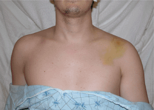 Figure 1.1. Clinical photograph showing swelling and ecchymosis over the left clavicle. |
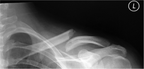 Figure 1.2. Preoperative AP radiograph demonstrating a midclavicular fracture with shortening and overriding of the fracture. |
Medial clavicular fractures, although rare, require special attention if any posterior displacement or intra-articular extension is found. Because it will provide the optimal visualization of the fracture and sternoclavicular joint, a computed tomography (CT) scan is often helpful when a complex, medial, clavicular fracture is present.
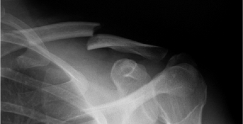 Figure 1.3. An apical oblique view helps minimize interference of the thoracic structures when viewing the fracture. |
Careful preoperative planning based on the fracture characteristics is essential to any orthopedic surgical procedure. Although the emphasis of this chapter is on plate fixation of acute clavicular fractures, other surgical procedures, with varying results, have been described. Intramedullary pins or nails are a viable treatment alternative because they allow for limited exposure and soft-tissue disruption. However, numerous case reports have been cited regarding pin migration out of the bone and into the lung (16), ascending aorta (17), pulmonary artery (18), abdominal aorta (19), and even the spinal canal (20). Although rare, the potential for migration still exists, even when precautionary measures such as using threaded pins or bending the pin at the end, are used.
Related posts:
 Radial Head Fractures: Open Reduction Internal Fixation
 Femoral Neck Fractures: Open Reduction Internal Fixation
 Femoral Shaft Fractures: Retrograde Nailing
 Proximal Tibia Fractures: Locked Plating
 Talus Fractures: Open Reduction Internal Fixation
 Open Reduction Internal Fixation Using an Extensile Lateral Approach for a Joint Depression Fracture
Stay updated, free articles. Join our Telegram channel

Full access? Get Clinical Tree


