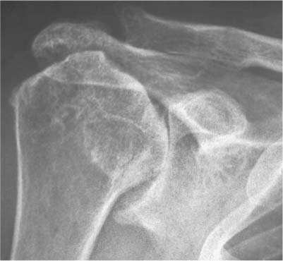
3 Classification of Rotator Cuff– Tear Arthropathy Cuff tear arthropathy (CTA) is not a unique pathologic entity. It is the common end stage result of several disease processes such as rheumatoid arthritis, rotator cuff (RC) tear arthropathy, or Milwaukee shoulder syndrome. The characteristic clinical and functional appearance of the common end stage of several disease processes is characterized as a painful arthritic shoulder with nonfunctional, irreparable cuff. By developing a substantial defect in the RC tendons, these disease processes lead to destabilization of the glenohumeral joint with subsequent superior migration of the humeral head and secondary severe damage to both the intraarticular and extraarticular elements. Massive RC defects lead to a loss of static or dynamic glenohumeral stabilization and to an anterosuperior displacement of the humeral head. The extent of displacement depends on the number and locations of tendons affected1–3 and their degree of involvement, the extent of atrophy of the muscles,2, 3 the structural integrity of the coracoacromial arch, and the extent and direction of the accompanying glenoid destruction. The consecutive anterosuperior displacement and instability of the humerus and the change in the center of rotation cause an insufficiency of the deltoid muscle.4 Biomechanical investigations done by Grammont5 and De Wilde6 have shown that a caudal and medial displacement of the glenohumeral center of rotation causes a significant increase in the moment of rotation of the deltoid muscle. Conversely, it could be assumed that the superior and lateral displacement of the center of rotation deteriorates the biomechanics of the deltoid. The previous classifications of Hamada and Fukuda18 or Favard19 did not have any therapeutic impact. They purely describe the natural course and explain the pathomorphologic consequences of large and massive cuff tears. Therefore, my group established a more functional and biomechanical classification of cuff tear arthropathies into four types focusing on the position and stability of the center of rotation on static (normal x-ray) and dynamic (fluoroscopy) radiologic investigation. We intended to develop a classification based upon treatment guidelines, and one based on, yet independent from, the underlying etiology. The RC tendons provide a major contribution to the dynamic stabilization of the glenohumeral joint by increasing the concavity-compression force in the joint.7,8 By their synchronous action, they oppose the displacing effect of the strong deltoid muscle, keeping the humeral head centered in the glenoid fossa throughout its movement.9 The coupled work of the infraspinatus and subscapularis tendons has been shown to be a major factor in superior glenohumeral stability, whereas the contribution of supraspinatus tendon is less significant.10–13 A massive tear, consisting of the supraspinatus tendon and at least one of the other RC tendons (in most cases the infraspinatus) makes the RC’s anterior and posterior force couple ineffective in both the vertical and the transverse planes. The result is a diminution of joint reaction force and a destabilization of the glenohumeral joint.11 In cases where the long head of biceps is still functional, it may oppose, to some extent, the superior migration of the humeral head.12 Nové-Josserand and colleagues1 demonstrated in a retrospective analysis on numerous (n = 246) patients with large and massive cuff tears that the additional involvement of the subscapularis tendon leads to a significant decrease of the acromiohumeral distance (an indicator of the superior migration of the center of rotation) in comparison to nonsubscapularis-involved two-tendon tears of the supraspinatus and infraspinatus tendons. The location of the defect, whether it is more a posterosuperior or an anterosuperior large or massive defect, is also important in relation to the amount of superior displacement. Posterosuperior defects have a bony buttress by the osseous arch of the acromion; therefore, the superior displacement has structural barriers and limits. Gagey36 described a biomechanically important tight fibrous frame consisting of collagen fiber bundles in the anterior part of the supraspinatus and in the superior part of the subscapularis, the biceps tendon and the coracohumeral ligament, which acts as a passive restraint against anterosuperior translation. Therefore, RC defects of the same size located in the anterosuperior section of the cuff leads to a greater amount of superior translation than posterosuperior defects. If the proximal pull of the deltoid is left unopposed, the humeral head migrates superiorly toward the coracoacromial arch. The deltoid, which has lost its fulcrum, is left with a smaller mechanical advantage and therefore must generate more force to perform its function. The humeral head then articulates with the coracoacromial arch superiorly and the superior glenoid rim inferiorly, leading to flattening of the superior part of the humeral head and tuberosities (“femoralization”), rounding and thinning of the coracoacromial arch (“acetabularization”) and destruction of the superior glenoid region (Fig. 3–1). The result is an incongruous, unstable joint with a higher joint friction and superiorly malpositioned center of rotation. The occurrence, expression, and presentation of the single morphologic features are multifactorial and mainly dependent of the underlying pathology and the pathomechanics of the RC tear. Otherwise, the pathomechanics of the RC tear is highly dependent on the size and location of the tear, the number of tendons involved, the integrity of the coracoacromial arch and the bony geometry of the glenoid. In contrast to the negative biomechanical effect of the superior migration of the center of rotation of the glenohumeral joint for the deltoid Grammont disclosed, 1 cm caudalization or medialization improves the deltoid-torque by 20 to 30%. In a recent study, De Wilde et al6 demonstrated in a computer model, that a simulated elongation of the deltoid along the humeral axis of ~10% with a stable center of rotation significantly improves the delta force especially in the critical 90-degree-abduction position. Although sharing a common functional result, it is important to recognize the various disease processes leading to CTA. The specific and characteristic parameters of the various processes greatly affect the time and aggressiveness of occurrence and the morphologic phenotypes of presentation of CTA. The different etiologies have a decisive influence on treatment and outcome.13 The most important etiopathologies are • Primary rotator cuff tear arthropathy14 • Post–rotator cuff–repair arthropathy • Inflammatory arthritis with extensive rotator cuff defect • Crystalline-induced arthritis arthropathy (Milwaukee shoulder)15 • Destructive arthritis • Primary osteoarthritis with extensive rotator cuff defect The characteristics of each etiopathology are discussed below. Figure 3–1 Typical x-ray of rotator cuff tear arthropathy showing superior migration, acetabularization, and superior glenoid erosion. CTA could be the result of a massive RC tear. The term introduced by Neer in 198314 refers to a primary massive RC tear that by virtue of mechanical superior instability and nutritional effects leads to a secondary glenohumeral joint destruction. The percentage of massive cuff tears that will end up as CTA is estimated to be between 0 to 25%, but it is very difficult to predict which massive tear will result in CTA.16 Post-CTA has similar pathoetiology and behavior as primary CTA. Rheumatoid arthritis (RA) is one of the most common causes of CTA. Between 48 to 65% of RA patients have significant glenohumeral joint involvement. About 24% of those having glenohumeral arthritis will have a simultaneous RC tear. The acromioclavicular joint is also frequently involved in the process, joining its cavity with that of the now joined synovial intraarticular and subacromial bursae spaces. Additionally, there are often severe osteopenia, erosions of the entire glenoid without osteophyte formation, and medialization of the glenohumeral joint.17 The Milwaukee shoulder syndrome was originally described by McCarty in 1981.15 This is an uncommon entity affecting shoulders of elderly people, predominantly women. It consists of a massive RC tear, joint instability, bony destruction, and large bloodstained joint effusion containing basic calcium phosphate crystals, detectable protease activity, and minimal inflammatory elements. Its relation to RC arthropathy is not clear and it might represent one spectrum of the above. The role of the basic calcium phosphate crystals in creating this syndrome is still controversial. Whether it is the cause of the articular damage through macrophage released proteases, or just the result of the osteoarthritic process is still unknown. Primary glenohumeral osteoarthritis is the most common reason for shoulder joint replacement; however, it is associated with RC tear in only 5% of patients, most of which are reparable. It is therefore uncommon for primary osteoarthritis to end up as CTA. The Hamada–Fukuda Classification18 is more or less a morphologic description of the natural course of massive RC tear and therefore only focusing on the group of (primary) CTA according to Neer.14
Biomechanics of Pathophysiology of Rotator Cuff Tears
Characteristics of Different Etiologies of Rotator Cuff Arthropathy
Primary Rotator Cuff Tear and Post– Rotator Cuff–Repair Arthropathy
Rheumatoid Arthritis
Crystalline-Induced Arthritis Arthropathy (Milwaukee Shoulder)
Primary Osteoarthritis with Extensive Rotator Cuff Defect
Classifications of Rotator Cuff Arthropathy
Hamada–Fukuda Classification
![]()
Stay updated, free articles. Join our Telegram channel

Full access? Get Clinical Tree


