Fig. 2.1
Seddon and Sunderland classification of nerve injury based upon histological neural changes
2.3.1.1 Neurapraxia
Neurapraxia is seen as motor paralysis, and it is the mildest injury type that is transient. There is no effect on nerve continuity. The transient nature of this injury is believed to be caused by a temporary disturbance in the conduction pathway that blocks neural transmission but does not damage the axon. Symptoms include motor paralysis (for motor nerves), numbness, tingling, and loss of vibration and postural sensation. All of these effects resemble the common effects of local anesthesia.
2.3.1.2 Axonotmesis
Axonotmesis occurs when there is complete interruption of the nerve fibers, but the connective tissues (endoneurium, perineurium, and epineurium) remain intact. It is a disturbance of nerve cell axon, with Wallerian degeneration occurring near the site of injury. This type of nerve injury is caused by a crush or pressure damage. Spontaneous regeneration is likely to occur following this type of injury [12]. The nerve as a mass is still in continuity [11].
2.3.1.3 Neurotmesis
Neurotmesis involves complete severance of the nerve. Functional loss is complete and recovery without surgical intervention is unlikely. There is a complete loss of motor and sensory function. If there is recovery, it is usually incomplete. It is important to note that clinically, there may no difference between axonotmesis and neurotmesis. Discerning the differences between the two entities includes:
1.
Prognosis: Axonotmesis may be expected to be followed by spontaneous regeneration, while in neurotmesis, signs of recovery fail to appear or may occur only following surgical repair [11] (Table 2.1).
Table 2.1
Neurosensory recovery based upon Sunderland classification
Sunderland | Recovery pattern | Rate of recovery | Need for surgery |
|---|---|---|---|
1st degree | Complete | Fast (days-weeks) | − |
2nd degree | Complete | Slow (weeks) | − |
3rd degree | Variable | Slow (weeks-months) | −/+ |
4th degree | Poor | Little/none | + |
5th degree | None | None | ++ |
2.
Time: The specific time frame for recovery differentiates between axonotmesis and neurotmesis. Axonotmesis may be followed by spontaneous recovery while neurotmesis is not. The drawback is that once a time limit, generally accepted as 90 days, has passed, the likelihood of spontaneous regeneration decreases significantly (9.3–62.9 %) [13].
3.
Exploration: The only precise and predictive method to differentiate between axonotmesis and neurotmesis. If performed in a conservative manner, exploration is the gold standard for distinguishing between the three basic classifications of nerve injury [11].
4.
Histology: It is important to emphasize that histological analysis is very reliable, however, clinically not useful.
In 1951, Sir Sydney Sunderland further stratified the three nerve injury types described by Seddon into five categories according to Wallerian axon degeneration and disruption of the endoneurial, perineurial, and epineurial tissues again based upon histological findings [14].
A first-degree injury is equivalent to Seddon’s neurapraxia and a second-degree injury is equal to axonotmesis. Third-degree nerve injury can be described as axonotmesis with endoneurial involvement. This category fits between Seddon’s pure axonotmesis and neurotmesis. Dependent upon the extent of the endoneurial damage, functional recovery may be possible. Sunderland further divides Seddon’s neurotmesis into fourth- and fifth-degree injuries. In a fourth-degree injury, all portions but the epineurium of the nerve are disrupted and surgery is necessary for treatment. Fifth-degree injury, similar to the classic neurotmesis, involves complete severance of the nerve including the epineurium [14].
In 1988, Mackinnon introduced a new injury pattern deemed useful to further classify nerve injuries. This classification scheme combines multiple types of nerve injuries seen in the Sunderland classification. It is in other words a mixed scheme where many types of nerve injury are combined and therefore there are variable degrees of recovery witnessed by the examiner and experienced by the patient. Electrodiagnostic tests are used to differentiate between first degree and other degrees of nerve injury; however, they will not differentiate between the recovery potential associated with each injury [15] (Table 2.2).
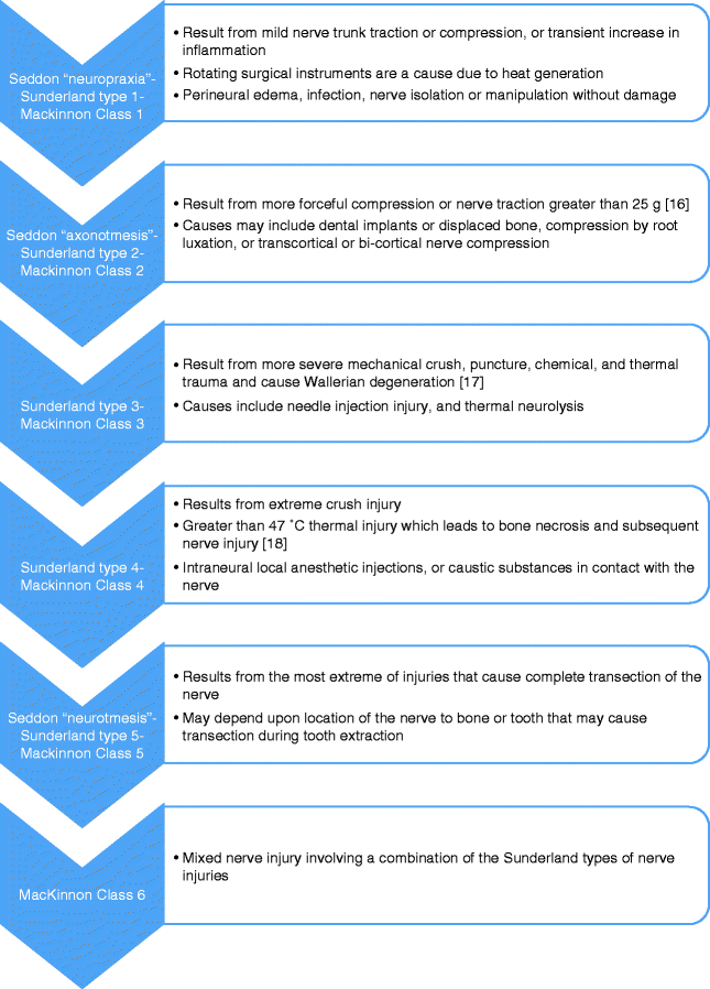
Table 2.2
Modified MRCS (Medical Research Council Scale)
Grade | Description |
|---|---|
S0 | No sensation |
S1 | Deep cutaneous pain in autonomous zone |
S2 | Some superficial pain and touch |
S2+ | Superficial pain and touch plus hyperesthesia |
S3 | Superficial pain and touch without hyperesthesia and static 2-point discrimination >15 mm |
Indicates USF (useful sensory function)a | |
S3+ | Same as S3 with good stimulus localization and static 2-point discrimination of 7–15 mm |
Indicates USFa | |
S4 | Same as S3 and static 2-point discrimination of 2–6 mm |
Indicates CSR (complete sensory recovery)a |
More recently, the modified Medical Research Council Scale (MRCS) has been applied to the classification schemes for trigeminal nerve injury (Fig. 2.2). The grading system provides a useful method to document and monitor neurosensory recovery, either spontaneously or following surgical repair.
2.4 Nerve Injury Classification via Independent Classification
Nerve injuries as discussed previously can be created by a myriad of causes, most notably, mechanical injuries such as that produced during third molar extractions, root canal instrumentation, dental implant placement, and osteotomy procedures.
Systemic processes may also influence nerve injury responses. These include chemical injury, infection, metabolic, genetic, and disease-related causes. These other mechanisms of nerve injury may be referred to as nerve classification independent.
2.4.1 Chemical Injuries
Nerve injury by chemicals has been noted in the literature from nerve agents used as chemical weapons to the common household bleach. Any chemical that can disrupt the conduction mechanism of the nerve can cause chemical nerve injury [19]. This discussion will be limited to chemicals that are used in dental and surgical settings.
Local anesthetics are commonly used in dentistry and oral and maxillofacial surgery, and it is generally considered a safe chemical to use in a healthy individual. Unfortunately, injection-related trigeminal nerve injury that can cause neurosensory disturbances (NSD) is a rare but present complication. The signs and symptoms associated with NSD are associated with a range including anesthesia, hypoesthesia, dysesthesia, allodynia, gustatory abnormalities, and spontaneous pain [20]. Estimates of the incidence of local anesthetic related NSD (temporary or permanent) in dental practice vary greatly, ranging from as high as 1:42 to as low as 1:750,000 disturbances per injection [20]. However, the true incidence remains elusive due the unknown number of unreported cases. Symptom resolution has been suggested to occur within 8 weeks; however, more longitudinal studies are still needed [21, 22]. The four major local anesthetics that were associated with the highest incidence of NSD were articaine 4 %, lidocaine 2 %, mepivacaine 3 %, and prilocaine 3 %, with articaine having the highest share of cases reported [20].
Other common chemicals that have been associated with chemical nerve injury are those used in root canal therapy during cleaning or sealing. Bleach, or sodium hypochlorite, is the most commonly used irrigant in endodontics due to its dual action as a powerful antimicrobial agent and its ability to dissolve organic soft tissue in the root canal system [23]. Bleach extravasation beyond the root canal apex into the surrounding tissue can cause a plethora of signs and symptoms ranging from weakness and paresthesia to neuropathic pain and tissue necrosis. The onset of these signs and symptoms can range from immediate onset to late onset [23]. All root canal sealants are neurotoxic to some degree depending upon the level of penetration of the epineurium. Unfortunately, dysesthesia can be the main NSD noted from sealant damage with a rate of 30 % [5].
2.4.2 Infection
A variety of bacteria and viruses can cause neural damage resulting in a condition known as peripheral neuropathy. The most notable virus to be discussed is herpes zoster virus causing postherpetic neuralgia (PHN) and Ramsey Hunt syndrome II (herpes zoster oticus). Herpes zoster has the highest incidence of all neurologic diseases. It occurs in approximately a half million individuals in the USA and has a lifetime occurrence of 20 % [24, 25]. After initial infection, the varicella-zoster virus establishes latency in the spinal and cranial nerve ganglia. After reactivation and replication, the viruses spread from the sensory nerve fibers in the ganglion to the involved dermatomes. Other than the dermatologic manifestations of the infection, there are a multitude of neurologic manifestations ranging from neuropathic pain to paresthesia [26]. The rash disappears within 2–4 weeks, but the most distressing symptom that may persist after resolution of the acute infection is pain, predominately allodynia. Pain that persists beyond 3 months is termed postherpetic neuralgia [27]. It is a pain that has been described using words such as stabbing, burning, gnawing, and shooting. The destruction of primary afferent C-fibers and resulting central hyperactivity is thought to be the cause of pain in PHN [28, 29].
Related posts:

Stay updated, free articles. Join our Telegram channel

Full access? Get Clinical Tree


