CHAPTER 2 Classical Patient Selection for Unicondylar Knee Arthroplasty
 The patient must be able to indicate the location of the knee pain along the medial joint line in the varus knee.
 The physical examination of the knee must confirm the location of the tenderness along the medial joint line with minimal to no tenderness in all other areas.
 The ACL should be intact to physical examination (the anterior drawer test should be negative even if the ACL is absent on an MRI examination).
Introduction
Unicondylar knee arthroplasty (UKA) has progressed through two separate time phases since the original designs were developed in the early 1970s. The first phase was fraught with problems related to the prosthetic designs and patient selection.1–4 The results were good to excellent for the first 10 years after the surgery in the hands of the designing surgeons. In the second decade the results did tend to taper off and were not as good as the reports of total knee arthroplasty (TKA).5,6 It was difficult for the standard orthopaedic surgeon to reproduce the findings of the designers, and interest decreased in the late 1980s and early 1990s. Insall’s data showed that only 6% of knees satisfied the criteria for UKA, and he favored TKA as the procedure of choice.7
Repicci introduced the limited surgical approach (minimally invasive surgery, or MIS) for UKA in the early 1990s, and interest in the procedure increased by the year 2000.8–13 Newer designs appeared, and the Oxford mobile-bearing UKA became very popular both in Europe and in the United States.14,15 With this new wave of interest, surgeons looked to improve the clinical results and reviewed the patient selection criteria, the surgical approach, and instruments. If the incorrect patient is chosen, the result will be compromised despite excellent surgical technique and prosthetic design. This chapter outlines the factors involved in the choice process that should lead to a more satisfactory overall result.
Physical Examination
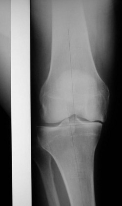
The examination should include inspection of the gait and, then, the full evaluation of both lower extremities. There should be a component of antalgia to the gait, and any thrust of the femur on the tibia through the stance phase should especially be noted. As the deformity progresses either in the varus or the valgus knee, the collateral ligament on the compressed side of the joint shortens and the ligament on the tension side lengthens. This ultimately leads to shifting of the tibia beneath the femur with impingement of the lateral tibial spine against the lateral femoral condyle in the varus knee and impingement of the medial tibial spine against the medial femoral condyle in the valgus knee (Fig. 2–1). The shift of the tibia correlates with a lateral thrust of the femur on the tibia through the stance phase of gait in the varus knee and a medial thrust in the valgus knee (Fig. 2–2). This finding is a relative contraindication to UKA and should alert the examiner to correlate the physical finding with the standing anteroposterior radiograph.
< div class='tao-gold-member'>
Stay updated, free articles. Join our Telegram channel

Full access? Get Clinical Tree


