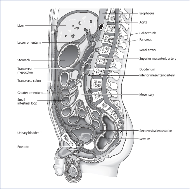
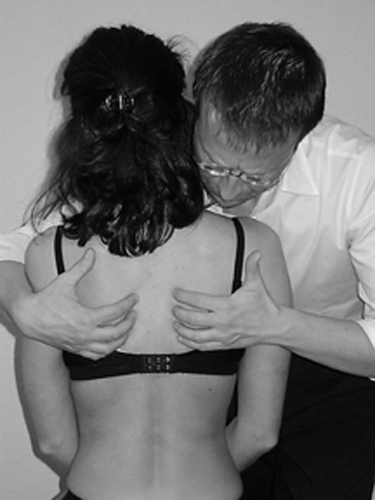
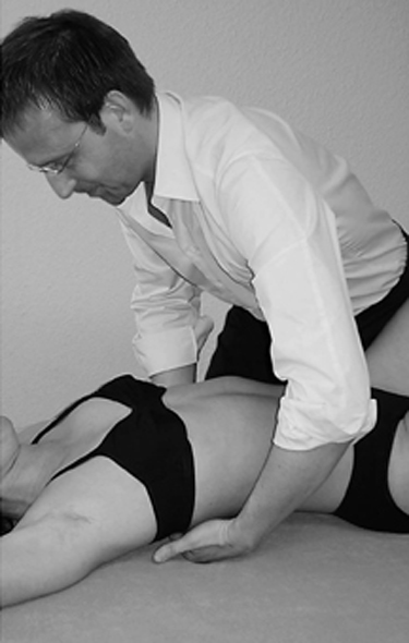
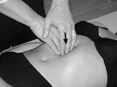
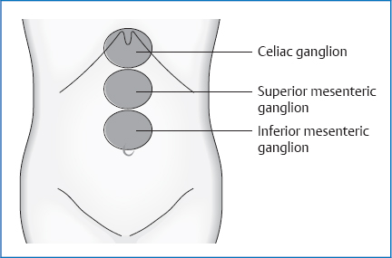
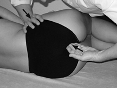
3 Circulatory Techniques according to Kuchera An organ can be influenced by its circulation, which includes the arterial, venous, and lymphatic systems, as well as the sympathetic and parasympathetic innervations. By means of the following treatment techniques, we can affect the trophic state of the organ. This is very important for organs that show pathology, e.g., gastritis. A prerequisite for these techniques is familiarity with circulatory anatomy, which is discussed in the chapters on individual organs. Fig. 3.1 The abdomen in sagittal section. The large vessel trunks for the abdominal space lie in front of the abdominal aorta and hence in front of the spinal column. Treating the spinal column (manipulation, mobilization, etc.) at the corresponding height stimulates the arterial supply of attached organs. The organs of the gastrointestinal tract drain their blood into the portal vein before it flows out through the liver into the inferior vena cava. Techniques that positively affect the portal vein, liver, or diaphragm improve venous drainage from the gastrointestinal tract. All techniques that promote lymphatic drainage improve the trophic state of the organ, e.g., diaphragm techniques, grand maneuver, etc. Parasympathetic. Techniques that treat the vagus or sacral parasympathetic nerve have a harmonizing effect on the internal organs, e.g., craniosacral techniques, treatment of the larynx, mediastinum techniques, etc. Sympathetic. On the basis of familiarity with the innervation of the organ, treatments that harmonize at the sympathetic level follow the course of the sympathetic nerves or plexus, e.g., sympathetic trunk stimulation by means of the rib-raise technique, diaphragm techniques, or stimulation of the prevertebral ganglia. Fig. 3.2 Starting Position The patient is in the supine position, legs stretched out, arms at the sides of the body. The practitioner stands to the side of the patient. Procedure Using the fingertips of both hands, make contact with the skin area lateral to the transverse process above the ribs. Place the fingers on both sides in such a way that the patient’s thorax is lifted passively off the table. Fig. 3.3 Treatment Maintain this position until fascial release takes place. Then shake the patient’s thorax rhythmically above the positioned fingers 8–10 times for sympathetic stimulation. Fig. 3.4 Fig. 3.5 Projection of the prevertebral plexus onto the abdominal wall. Starting Position The patient is in the supine position. The practitioner stands next to the patient. Procedure At the height of the projection of the preaortic plexus on the abdominal wall, place the fingers of both hands next to each other in the center line and let them sink into the depth of the abdomen until you reach the plexus. It may be necessary to stop repeatedly on the way in and await fascial release. Treatment When you have arrived at the plexus, maintain pressure until you have obtained fascial release. Then stimulate the preaortic plexus by means of repeated rebounds. Fig. 3.6 Starting Position The patient is in the lateral position, hip and knees bent to 90°; the side to be treated faces up. The practitioner stands behind the patient. Procedure Place the fingers of the caudal hand medial to the ischiadic tuber and lateral to the coccyx, near the tuber, with the palm facing up. The cranial hand grips the iliac crest from anterior near the anterosuperior iliac spine (ASIS). Treatment The caudal hand exerts careful pressure cranially and anteriorly (in the approximate direction of the cranial hand). Wait for fascial release and then increase the pressure. The pressure of the caudal hand is applied in the direction of the greatest fascial tension. The cranial hand maintains counterpressure. You can work with careful vibrations.
Objective
Principle of the Techniques
Arterial Stimulation
Venous Stimulation
Lymphatic Stimulation
Vegetative Harmonization
Techniques
Vegetative Harmonization
Rib-Raise Technique
Treatment of the Preaortic Plexus
Treatment of the Ischiorectal Fossa
Related posts:


Stay updated, free articles. Join our Telegram channel

Full access? Get Clinical Tree


