2 Circulatory physiology
2.1 Circulatory function – generalities
2.1.1 Conceptualization
One can simplify the workings of the circulatory system by looking at functional parts:
• An alternating pump: the heart
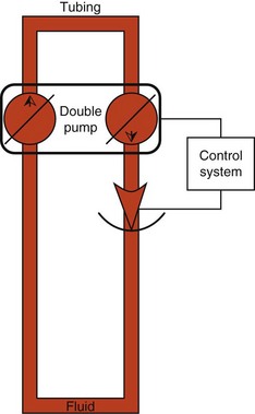
• A hydraulic circuit: the vascular system, which can be considered, at least in the short and middle term (independent of renal resorption) a constant volume. This hydraulic circuit (Fig. 2.1) comprises:
• the cardiovascular system has some porous tubes (capillaries)
• the cardiovascular system is made up of elastic tubular elements and not rigid structures
• the volume of its contents is subject to changes depending on digestive absorption, as well as renal and digestive elimination.
These characteristics fundamentally complicate any more basic conceptualization.
2.1.2 Pressures
Arterial pressure
• increases during the emptying phase (ventricular systole)
• decreases during the filling phase (ventricular diastole).
Average blood pressure
This can be expressed by the formula:
For example, in a person whose arterial pressure is 140/80 mmHg, the average pressure is:
Values
Arterial pressure varies with:
• Sex: up to the age of 40 years males have higher arterial values than women, becoming lower than women after age 50 (D’Alché 2008).
• Age: on average blood pressure rises with age.
• Cardiac output: a rise in output tends to increase blood pressure. For example, during physical exertion, arterial pressure increases. By contrast, blood pressure falls during sleep. Systolic pressure comes down as much as 10 to 30 mmHg, and diastolic pressure lowers 5 to 10 mmHg. (D’Alché 2008).
• Blood volume: increased blood volume increases blood pressure. An injection of 250 mL water causes blood pressure to rise by 10 mmHg within 60 min.
• Blood flow to individual organs depends on the degree of vasoconstriction of the arteries supplying the particular organ. Digestion modifies arterial pressure.
2.1.3 Arterial tension
In France, the term ‘arterial tension’ refers to arterial pressure measurement.
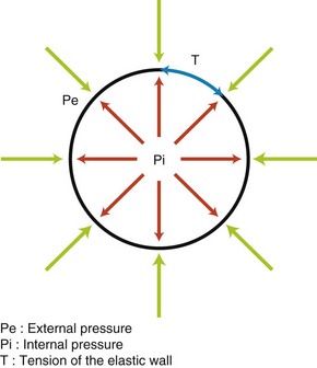
Normally, the term fluid pressure denotes fluid pressure on the walls of a deformable tube. Blood vessels are elastic tubes and as such are deformable. Laplace’s law expresses the relationship between the tension, T, exerted on the wall of such a tube and the prevailing pressure at the interior of the tube (Fig. 2.2).
2.2 Cardiac physiology
2.2.1 Cardiac mass
Cardiac cycle
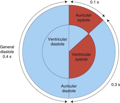
The contraction of the heart, or systole, is followed by a period of rest, called diastole. Together they constitute the cardiac cycle (Fig. 2.3).
The resting time between heart beats is about twice that of a contraction.
Terminology
Although they are brief events in the cardiac cycle, diastole and systole are of a particular duration nonetheless. Sometimes it is necessary to be more specific about a particular moment during these occurrences or, in contrast, it can be helpful to refer to the overall picture. Table 2.1 summarizes the meaning of these various prefixes.
Cardiac cycle
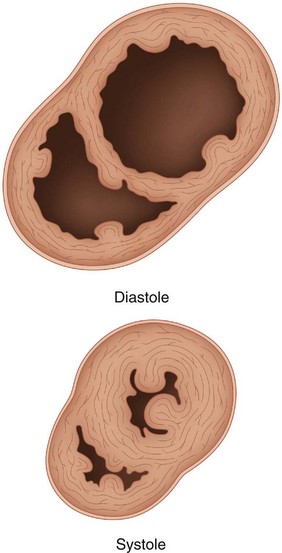
The heart can be likened to a hollow muscle that propels blood by alternately contracting and relaxing. This sequence affects the size of various cavities, especially the ventricles. During ventricular contraction (systole) the walls thicken and the cavity size diminishes, whereas during ventricular relaxation the walls thin and the cavity enlarges (Fig. 2.4).
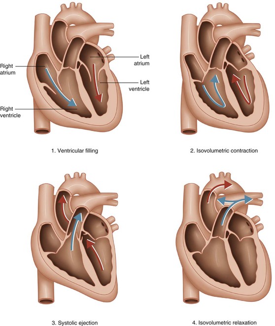
The adult human heart ‘beats’ about 70 times per minute, which means the four action phases of the heart are accomplished in less than 1 s. The four phases (Fig. 2.5) are:
• The ventricular filling phase, during which the atrioventricular valve is open. Blood flows from the atrium towards the ventricle, while the aortic valve (for the left ventricle) and the pulmonary valve (for the right ventricle) remain closed. During this phase, blood flow first flows passively from the atrium to the ventricle, and then more actively during auricular diastole.
• In the isovolumetric contraction phase, myocardial ventricular contraction increases the pressure in the ventricle, and all valves are closed.
• Systolic ejection begins when pressure in the ventricle (ventricular pressure) overtakes the pressure in the aorta (aortic pressure) or the pulmonary artery, during the course of which the blood is expelled from the ventricle. The semilunar valves are opened.
• Isovolumetric relaxation begins at the end of the ventricular contraction. Ventricular pressure falls below that of the aorta and the pulmonary artery causing closure of the semilunar valves. The atrioventricular valve remains closed.
2.2.2 Cardiac output
Volume of systolic ejection
1 The pre-charge corresponds to the volume of blood present in the left ventricle just before ejection. Volume depends on the pressure of venous return to the heart, called central venous pressure (CVP).
2 The contractility of the myocardium, regulated by the autonomic nervous system.
3 The post-charge represents those factors that oppose the work of the heart:
Stay updated, free articles. Join our Telegram channel

Full access? Get Clinical Tree


