11 Children’s fractures
Cases relevant to this chapter
Essential facts
1. Children’s bones have a lower modulus of elasticity than adult bones and are more susceptible to bending forces.
2. Children’s fractures are more likely to occur in the metaphysis, where there is less cortex than medulla, compared to the diaphysis, which has an equal amount of cortex and medulla.
3. Fracture patterns in children are as follows: plastic deformation, buckle or torus fracture, greenstick fracture, complete fracture and physeal (growth plate) injury.
4. Fracture healing in children is more rapid than in adults; in addition there is more capacity to remodel, so that some residual angulation or translation after reduction may be acceptable.
5. Physical abuse is the most common category of non-accidental injury and is characterized by bruises, lacerations, thermal injuries and fractures (which have a prevalence of 11–55% in these children).
6. The following features suggest non-accidental injury: a vague history; inconsistency with clinical findings or child’s developmental stage; unexplained delays in seeking help; evasive or aggressive parents or carers; the presence of other injuries; and previous concerns regarding the child or sibling(s).
Structure of a child’s bone
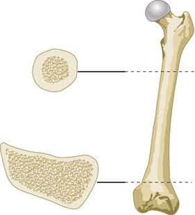
A cross-section of the diaphysis and metaphysis of a child’s long bone will show that the diaphysis is approximately circular and there is also an approximately equal amount of cortex and medulla. On the other hand, the metaphyseal region is generally oval in cross-section and there is relatively less cortex with respect to the medulla. Figure 11.1 illustrates this using the femur as an example. If forces are applied to the bone it is more likely to fail in the metaphyseal region and this accounts for the large number of metaphyseal injuries of long bones seen after low-velocity trauma in children. A greater amount of energy is required to fracture the diaphysis (Fig. 11.2).
Types of fracture
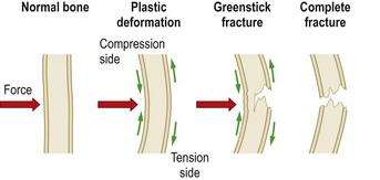
There are five distinct fracture patterns in children (Box 11.1). The first four will be considered here and physeal injuries in the section on growth plate injuries. The fracture patterns result from the direction and amount of energy that has been applied to the bone (see Fig. 11.2). Initially the bone will bend, plastic deformation occurs as a result of micro-fractures of the cortex and the bone will not return to its original shape. This pattern is not usually seen in adults. It is seen most often in the forearm and leg where plastic bowing of the ulna or fibula may accompany a radial or tibial fracture. With an increasing amount of energy the bone will start to fail and buckle. Eventually the bone will fail on the tension side whilst the compression side will continue to bend, thus producing the characteristic greenstick fracture (Fig. 11.3). More energy applied to the bone will cause a complete fracture of the compression side.
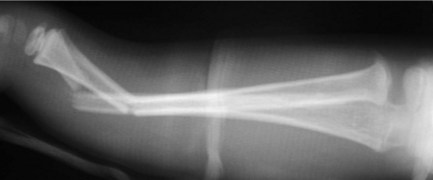
FIGURE 11.3 Greenstick fracture of the radius (there is one intact cortex) and complete fracture of the ulna
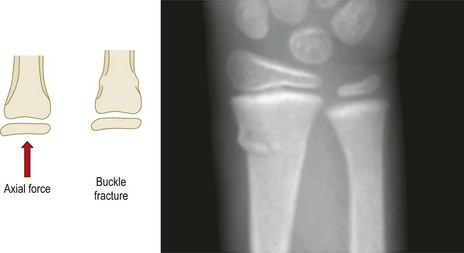
An axial or longitudinal force applied to a long bone in a child may produce buckling of the metaphysis. This type of fracture is also known as a torus fracture (Fig. 11.4). Torus is the name given to the band around the base of a Grecian column and in geometry represents the shape of a doughnut.
In a complete fracture three anatomical patterns are recognized (Box 11.2). These may also be comminuted (those containing more fragments of bone other than the proximal and distal components) or segmental (the same bone has been broken at two distinct sites).
Spiral fractures are produced as a result of rotational forces being applied to the bone; they are usually low-velocity injuries and may arise where one part of a limb is fixed relative to another. Oblique fractures are unstable and arise as a result of an increasing amount of force being applied to the bone. Transverse fractures tend to occur as a result of three-point bending. Comminuted and segmental fractures are a feature of high-velocity trauma (Fig. 11.5).
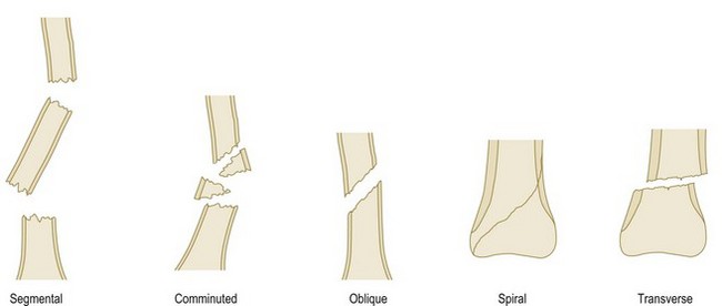
FIGURE 11.5 Shows the three patterns of a complete fracture, which may also be comminuted or segmental
Physeal (growth plate) injuries
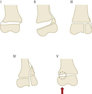
Children are unique in having growth plates, and the growth plate is thought to be an area of relative mechanical weakness compared with the bone of the metaphysis and epiphysis. Physes are more susceptible to injury from rotational forces rather than angulation or traction. The commonly used classification of physeal injuries by Salter and Harris is illustrated in Figure 11.6. The type I injury is a fracture through the physis without involving the bone of the metaphysis or epiphysis. The type II injury is the commonest and carries the best prognosis. Here the fracture propagates through the physis and includes a triangular portion of the metaphysis (always on the compression side of the fracture where periosteum is intact). The type III fracture passes through part of the physis and then across the epiphysis, whereas the type IV passes across the epiphysis, physis and metaphysis. Both type III and IV injuries involve the articular surface of the joint as well as the physeal plate and should be reduced anatomically, which often entails open reduction and internal fixation. The type III and IV injuries generally carry a worse prognosis because of the likelihood of physeal growth arrest from the disruption to the physis and the risk of post-traumatic arthritis of the joint if the articular surface is not reduced accurately. A growth arrest may cause an angular deformity because the uninjured part of the physeal plate continues to grow whilst the arrested side does not (Fig. 11.7). Type V is a compression injury to the physis and, like type I injuries, it does not involve the bone of the metaphysis or epiphysis. The injury may be diagnosed only in retrospect if a partial growth arrest of the plate becomes evident later.
< div class='tao-gold-member'>
Stay updated, free articles. Join our Telegram channel

Full access? Get Clinical Tree


