Chest Pain (Case 4)

Chapter 8
Chest Pain (Case 4)


Arzhang Fallahi MD and Michael Kim MD


Case: A 54-year-old man with a history of smoking, hypertension, and hyperlipidemia comes to the emergency department complaining of chest pain. He has had hypertension for 20 years but is poorly compliant with his antihypertensive regimen. He complains of chest pain while exercising and can make it up only two flights of stairs before having to rest. The day of presentation, he was walking up the stairs when he noticed a sudden onset of chest pressure radiating down the left arm associated with diaphoresis, shortness of breath, and nausea. The patient forgot to bring in his medications but says he is not taking what he was given, which was hydrochlorothiazide 25 mg daily, atorvastatin 20 mg daily, and aspirin 81 mg daily. He has a family history of hypertension, and his father died at the age of 45 from a myocardial infarction (MI). On examination he is a diaphoretic man weighing 150 kg, his pulse is 110 beats per minute (bpm), and his blood pressure (BP) is 170/95 mm Hg. His exam is notable for elevated neck veins, coarse breath sounds bilaterally, an S3 gallop, and 1+ edema in his lower extremities.


Differential Diagnosis*


images


*The differential diagnosis of chest pain is quite broad; this box is limited to the causes elucidated in the Clinical Entities section.


Speaking Intelligently


The assessment when approaching a patient with chest pain is as follows:


1. Assess airway, breathing, and circulation with intravenous access, oxygen administration, and cardiac monitoring, with initial blood work (including serum troponin).


2. Although acute coronary syndromes represent the leading cause of death in adults in developed countries, one should quickly assess and consider other immediately life-threatening conditions such as aortic dissection, pulmonary embolism, tension pneumothorax, pericardial tamponade, and mediastinitis (e.g., secondary to esophageal rupture).


3. Characterize the chest pain: onset (abrupt, gradual), position (localized to small area, diffuse), quality (tight/pressure, burning, fullness, knot, Levine sign [where the patient places his or her fist in center of chest]), radiation (to neck, throat, lower jaw, teeth, upper extremity, or shoulder), aggravating factors (association with eating, exertion or stress induced, positional, worse with cough or deep breathing), alleviating factors (relieved by antacids, nitroglycerin, or rest), associated symptoms (belching, vomiting, diaphoresis, dyspnea, cough, syncope, palpitations, underlying psychiatric disorders such as anxiety, depression, somatization).


4. Risk-stratify patients for coronary heart disease (Framingham: age, gender, high-density lipoprotein cholesterol [HDL], low-density lipoprotein cholesterol [LDL], systolic blood pressure, diabetes, smoking), as well as assessing thrombolysis in MI (TIMI) risk score, which categorizes a patient’s risk of death and ischemic events, providing a basis for therapeutic decision making.


5. Depending on the patient’s presentation, comorbidities, risk factors, and initial assessment, therapy can be conservative or invasive.


6. Quick and thorough assessment of the patient is of utmost importance. Often with acute coronary syndromes, the more time that passes the more myocardium is jeopardized. Remember that “time is myocardium.”


PATIENT CARE


Clinical Thinking


• First and foremost, the patient must be quickly assessed systematically for airway, breathing, and circulation. Prompt intravenous access, administration of oxygen, and cardiac monitoring should be instituted. Always consider the immediate life-threatening causes of chest pain: MI, aortic dissection, pulmonary embolism, tension pneumothorax, pericardial tamponade, and mediastinitis (e.g., secondary to esophageal rupture).


• Given the clinical presentation, this patient’s chest pain is most likely cardiac in nature. Angina can be broken down into two main subtypes: stable and unstable. Stable angina is defined as chest discomfort that occurs with exertion or stress and that is relieved by rest or nitroglycerin. Unstable angina is a type of acute coronary syndrome that encompasses myriad conditions including new-onset chest pain, rest angina, accelerating pattern of previously stable angina, post–myocardial infarction angina, and angina after a revascularization procedure.


• This patient’s presentation is classic for an acute coronary syndrome (ACS), which can be divided into three main subtypes: ST-elevation MI (STEMI), non–ST-elevation MI (NSTEMI), and unstable angina (UA). Three primary presentations that suggest ACS are (1) rest angina usually lasting more than 20 minutes, (2) new-onset angina that significantly limits physical activity, and (3) increasing angina that is more frequent, is longer in duration, and occurs with less exertion that previous angina. UA can present as new angina, rest angina, early post-MI angina (chest pain occurring within 48 hours after an acute MI), and post-revascularization angina. UA and NSTEMI are often indistinguishable on initial evaluation, as elevation of serum troponins and/or creatine kinase–isoenzyme B (CK-MB) is needed to distinguish the two and takes as long as 4 to 6 hours post MI to show elevations. UA and NSTEMI differ in whether ischemia is severe enough to cause enough myocardial damage to release markers of myocardial injury. NSTEMI often shows ST-segment depression (defined for acute MI as new horizontal or down-sloping ST depression greater than or equal to 0.05 mV in two contiguous leads and/or T inversion greater than or equal to 0.1 mV in two contiguous leads with prominent R-wave or R/S ratio greater than 1). STEMI is defined as ST-segment elevation with serum cardiac biomarker elevation. On electrocardiogram (ECG), acute ST-elevation MI is defined as elevation at the J-point in two contiguous leads with cutoff points: greater than or equal to 0.2 mV in men or greater than or equal to 0.15 mV in women in leads V2 to V3 and/or greater than or equal to 0.1 mV in other leads.


• Each of the three subtypes of ACS has a different therapeutic management algorithm. If considering an invasive strategy, prompt communication with the cardiology team and catheterization laboratory is in order. Early initial assessment of the patient is critical to provide timely care regardless of an invasive or conservative approach.


History


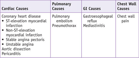
The history should be focused on risk stratification for cardiac disease and nature of the chest pain to classify as cardiac or noncardiac. In an acute setting, the history must be focused to rule out the most life-threatening conditions.


• The nature of chest pain is important in determining if the pain is cardiac or noncardiac in origin. Determine the onset, position, quality, radiation, severity, timing, aggravating factors, alleviating factors, and associated symptoms. Risk stratification is also helpful; evaluate such factors as patient age, gender, HDL, LDL, systolic blood pressure, diabetes, and smoking. Determine if the patient has had an MI before or chest pain in the past.


• Medication, diet, and social history: Is the patient taking antihypertensive medications or lipid-lowering agents? Is the patient using sublingual nitroglycerin? Does the patient use illicit drugs? Sympathomimetic agents such as cocaine may cause chest pain or MI, so their involvement is important to know for management.


• Family history: Early-onset coronary artery disease (CAD) is defined as CAD occurring in men younger than 55 years and women younger than 65 years.


• Other comorbidities: peripheral vascular disease, obesity, and renal disease, which can increase risk for CAD; recent stroke or intracranial hemorrhage.


Physical Examination


• A quick, but focused, physical exam should be done in all patients with chest pain, as it can give clues to the causes and effects of the chest pain.


Vital signs: Assess the patient for heart rate, hypertension, or hypotension (assess if patient appears to be in cardiogenic shock or in hypertensive crisis). The patient should also be assessed for respiratory distress and oxygen saturation.


General appearance: How does the patient appear? Look for diaphoresis, respiratory distress, and mottled appearance of skin indicative of poor perfusion.


Funduscopic exam: While most often difficult in an acute setting, a good funduscopic exam can show degree of hypertensive retinopathy; in cases where history is limited, this can provide evidence of poorly controlled blood pressure.


Respiratory exam: Make sure the trachea is in the midline to rule out a tension pneumothorax, listen to breath sounds bilaterally to make sure they are symmetric and to listen for any signs of fluid accumulation either from pulmonary and/or cardiac dysfunction or from other disease. Percussion may help identify areas of consolidation.


Cardiac auscultation: Determine whether the heart rate is regular or if there could be an underlying dysrhythmia. Determine whether the heart sounds seem distant, which may suggest a pericardial effusion; assess for murmurs and abnormal heart sounds (S3 gallop may indicate congestive heart failure or a dilated chamber, while an S4 gallop may indicate the stiff left ventricle of hypertensive heart disease). Look at neck veins to see if they are distended, which may indicate overload or inability to adequately distribute blood volume.


Vascular auscultation (carotid bruits): to assess atherosclerotic status.


Abdominal examination: Assess to make sure there is no evidence of esophageal rupture, acute abdomen, or possible gastrointestinal bleeding, which could have led to the ACS.


Neurologic examination: Quickly assess patient’s mental status to determine if there is adequate perfusion to the brain. A screening neurologic exam should be performed to see if there are any focal deficits that might preclude use of thrombolytic therapy.


Extremities: Check to see if extremities are warm to assess perfusion. If there is a discrepancy between lower and upper extremities, be sure to consider other vascular causes such as thrombosis or dissection.


Tests for Consideration


ECG is critical to obtain in all patients with chest pain.
ECG can be used to determine if ST elevations are present or if there are T-wave depressions indicative of ischemia.
Diffuse ST elevations may indicate a pericarditis.
Dysrhythmias may also be identified. Presence of a bundle branch block may mask an ST-elevation MI.


$27


Cardiac markers: Serum troponins and/or CK-MB levels are needed to distinguish unstable angina from the other two types of ACS. These are markers of myocardial injury significant enough to result in release of enzymes. More than one set of enzymes should be tested, as they take as long as 4 to 6 hours post MI to show elevations.


$49


Metabolic panel: Serum creatinine and electrolyte tests will help determine if an underlying metabolic disturbance is resulting in cardiac dysfunction. Severe hyperkalemia can lead to fatal dysrhythmias. Particular attention should be given to potassium and magnesium, as low levels can result in a higher rate of ventricular fibrillation in ACS. Creatinine level assessment is helpful to assess renal function and whether the kidneys are being perfused.


$12


Complete blood count: It is important to determine if the patient has an underlying infection (suggested by an elevated white blood cell count), which may be contributing to ACS.
Hemoglobin and hematocrit are important to determine if the patient is anemic, which could lead to cardiac ischemia as well as indicate a potential hemorrhage.


$11


Liver function tests may be helpful to ascertain if there is a gastrointestinal cause of the chest pain.


$12


Coagulation tests are helpful to determine if the patient is prone to thrombosis or if the patient is at high risk of bleeding. They are also helpful if the patient is to undergo an invasive procedure.


$15


IMAGING CONSIDERATIONS


→ Chest radiography is helpful to assess if any pulmonary disease or process such as a pneumonia, effusion, or pneumothorax is present. Cardiomegaly can be assessed but poorly so with a portable chest radiograph often obtained in an emergency situation. This can also help determine whether there is a widened mediastinum, which may be suggestive of an aortic dissection.


$45


→ Echocardiography is typically not used to evaluate chest pain but is used when a noncardiac cause such as aortic dissection, pulmonary embolus, pericarditis, or pericardial effusion is suspected. It also can be used to visualize wall motion abnormalities within seconds of coronary artery occlusion.


$393


→ Nuclear imaging with thallium-201 and technetium-99m sestamibi is helpful in certain clinical situations, as these agents accumulate proportional to myocardial perfusion. The 2003 joint task force of the American College of Cardiology (ACC), American Heart Association (AHA), and American Society for Nuclear Cardiology gave a class I indication in patients with suspected ACS where initial serum cardiac markers and ECG are nondiagnostic.


$300


→ CT. While not routinely used initially, it may be of some use in patients at low risk for ACS. Patients can also be scanned using a “triple rule-out” algorithm, which aims to assess for aortic dissection, coronary disease, pulmonary embolism, and other thoracic diseases.


$262

Stay updated, free articles. Join our Telegram channel

Oct 3, 2016 | Posted by in MANUAL THERAPIST | Comments Off on Chest Pain (Case 4)

Full access? Get Clinical Tree

Get Clinical Tree app for offline access