FIGURE 6.48 Dorsal aspect of the right hand. (From Tank PW, Gest TR. Lippincott Williams & Wilkins Atlas of Anatomy. Philadelphia, PA: Lippincott Williams & Wilkins, 2009.)
PATIENT POSITION
- Supine on the examination table with the head of the bed elevated 30 degrees.
- The affected wrist is held in a neutral position between supination and pronation.
- The wrist is supported with the placement of chucks pads or towels.
- Rotate the patient’s head away from the side that is being injected. This minimizes anxiety and pain perception.
LANDMARKS
1. With the patient supine on the examination table, the clinician stands lateral to the affected hand.
2. Locate the CMC joint by palpating the thumb metacarpal bone in a distal-to-proximal direction. At the proximal aspect of the first metacarpal, there will be tenderness as the examiner’s finger passes over, then drops into the CMC joint. This is located between the first metacarpal and the trapezium bone. The patient will report tenderness in this joint.
3. Mark the injection point directly over the CMC joint.
4. At that site, press firmly on the skin with the retracted tip of a ballpoint pen. This indention represents the entry point for the needle.
5. After the landmarks are identified, the patient should not move the hand or thumb.
ANESTHESIA
- Local anesthesia of the skin using topical vapocoolant spray.
EQUIPMENT
- 3-mL syringe
- 25-gauge, 5/8 in. needle
- 0.25 mL of 1% lidocaine without epinephrine
- 0.25 mL of the steroid solution (10 mg of triamcinolone acetonide)
- One alcohol prep pad
- Two povidone–iodine prep pads
- Sterile gauze pads
- Sterile adhesive bandage
- Nonsterile, clean chucks pad
TECHNIQUE
1. Prep the insertion site with alcohol followed by the povidone–iodine pads.
2. Achieve good local anesthesia by using topical vapocoolant spray.
3. Position the needle and syringe perpendicular to the skin with the needle tip directed posteriorly toward the first CMC joint.
4. Using the no-touch technique, introduce the needle at the insertion site (Fig. 6.49).
5. Advance the needle down into the joint.
6. Inject the steroid solution as a bolus into the joint. The injected solution should flow smoothly into the space. If increased resistance is encountered, advance or withdraw the needle slightly before attempting further injection.
7. Following injection of the corticosteroid solution, withdraw the needle.
8. Apply a sterile adhesive bandage.
9. Instruct the patient to move his or her thumb through its full range of motion. This movement distributes the steroid solution throughout the CMC joint.
10. Reexamine the CMC joint in 5 min to confirm pain relief.
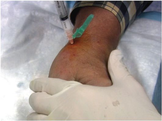
FIGURE 6.49 Left hand—thumb CMC joint injection.


