Fig. 7.1
Low grade chondrosarcoma of the pelvis
Anatomy
The pelvic bones have a complex anatomy both in their three-dimensional structure and their relationship to one another. The pelvis forms a ring, therefore encompasses a full 360°. The pelvis is also relatively inaccessible, particularly when compared to the long bones, and it is large. Therefore pelvic surgery often involves moving the patient intraoperatively to gain access. This coupled with the complex multiplanar structure makes pelvic surgery very challenging.
Consistency
Bone malignancies have variable consitencies, but often have cystic elements which may burst during the removal of the tumor. The tumor is also weaker than the surrounding healthy bone and may fracture upon removal. This makes it more difficult for the surgeon to remove the entire tumor en bloc and avoid tumor spill.
Margins
In order to reduce the risk of recurrence, it is accepted practice throughout oncology to achieve negative but also wide resection margins; that is, the tumor is removed with a surrounding margin of healthy tissue to ensure the entire tumor has been excised. This is very difficult to achieve during pelvic bone tumor surgery due to the size of the tumors being removed, difficulty of access, close proximity of vital structures and the multiplanar complexity of the structures involved. In addition, the late presentation of these tumors often allows the tumor to have invaded into local structures such as the pelvic veins or organs. Figure 7.2 demonstrates a pelvic chondrosarcoma in close proximity to the bladder, but with a clear plane for resection, Fig. 7.2a demonstrates a tumor invading into the bladder wall. Wide excision would be impossible in the case in Fig. 7.2a without a partial cystectomy. Additional difficulty occurs when a tumor has close anatomical relations to joints. It is common practice to preserve the joint architecture and articular surfaces during surgery to provide a better functional result, but often this is hampered by the desire to achieve a wide excision margin. Figure 7.3 shows intra-lesional, marginal, wide and radical resection margins. In patients with musculoskeletal malignancy the ultimate aim is to perform a wide-local resection and achieve adequate disease-free margins. Inadequate resection margins (intra-lesional or marginal) are frequently obtained [13]. The importance of achieving adequate surgical margins with these tumors is highlighted by the fact that local recurrence rates of up to 70 %and 92 % have been reported for pelvic tumors following marginal and intra-lesional resections respectively [13, 14].
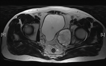
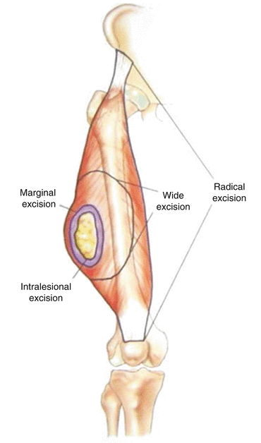
Fig. 7.2
Axial MRI scan of a pelvic chondrosarcoma with the cystic area and very thin soft tissue margin between the tumor and bladder
Fig. 7.3
Diagram depicting resection margins. In pelvic bone tumors, wide excisions are required to decrease recurrence rates
Reconstruction
Unlike the long bones, it is impossible to design a one-design-fits-all prosthesis to implant following pelvic tumor resection. In order to achieve a successful reconstruction, the prosthesis must fit the resection margins exactly to preserve the mechanics of the pelvic girdle. Debate rages as to whether the pelvic ring needs closing by reconstruction with most surgeons favoring not to close the ring. The complexity of the pelvis makes designing and fitting functional reconstructions that are durable and allow patients maximum function incredibly challenging. Poorly fitting prostheses result in damage to existing healthy bone and revision surgery, which is all the more complex for the deranged anatomy caused by the original tumor and primary surgical procedure. The long term survival of the reconstruction by endoprostheses is 75 % [15] and 85 % for massive allograft reconstruction [16].
Complications
Patients with malignancies of the pelvis are at a higher risk of treatment failure than other patients with similar tumors located in a limb [17]. Treatment failure can include recurrence, prosthesis failure, amputation and nerve damage. All of these problems stem from the difficulty in achieving adequate resection margins, and difficulty with reconstruction and large dead space. All major series have reported complication rates of in excess of 50 % following reconstruction. The complication rates following resection without reconstruction are lower and may still produce good functional results.
Function
The primary aim of pelvic tumor surgery is to remove the tumor completely to prevent recurrence. However, a key secondary aim is the need to preserve the function of the patient as much as possible. The functions to keep in mind are; transfer of weight from the upper axial skeleton to the lower limbs, especially during movement; providing attachment for muscles and ligaments used in locomotion; protecting the abdominal and pelvic viscera.
Suitability of the Pelvis for Navigation
The pelvis particularly lends itself to computer-assisted surgery as it has multiple easily identifiable bony prominences to use as reference points. Accurate registration is important as it allows the computer to build up a picture of the patient’s anatomy in space and therefore allows for direct correlation between the two-dimensional imaging studies and the three-dimensional surgical field by point to point and surface matching. This facilitates accurate orientation, tumor location and reconstruction thereby reducing the surgeon’s margin for error. The anterior superior iliac spines (ASIS), anterior inferior iliac spines (AIIS), posterior superior iliac spines (PSIS), the top of the iliac notch or any other easily identifiable anatomical landmarks can be used for registration. The registration error should be <1 mm before proceeding with resection.
However, where the shape of the pelvis aids registration, it hinders the practicality of performing the surgery. Pelvic tumors are often very large and may require resection in multiple planes due to the unique geometry of the pelvis. There are also many critical structures that must be avoided during pelvic surgery, such as the sciatic nerve and the iliac vessels as they pass through the sciatic notch, the bladder and the peritoneum. The use of navigation can significantly reduce the risk of damage to vital structures by allowing the surgeon to know their location relative to the osteotome, as well of the depth of penetration of the instruments. This is particularly useful in the sacrum, where uninvolved sacral nerve routes can often be spared, improving the patient’s neurological outcome after surgery.
Evolution of Surgical Techniques
All surgery requires extensive planning with knowledge of the patient’s and the tumor’s anatomy to enable a suitable implant to be designed. Since the advent of CT and MRI scanning, incredibly detailed three-dimensional representations of the tumor and surrounding anatomy can be isolated and explored before the operation. However, translating this information from view screen to intraoperative field can be difficult [17], resulting in inadequate resection margins or excessive removal of healthy tissue. Both of these scenarios result in unfavorable outcomes for patients. Inadequate resection margins (intra-lesional and marginal) frequently lead to local recurrence [14]. Excessive removal of bone causes difficulties to arise when trying to fit the implant or allograft. If this is not accurately done, there is a risk of non-union, disrupted biomechanics and implant failure.
Conventional techniques involve resection and reconstruction done ‘freehand’, with the scans available for reference. This has been shown to be highly inaccurate in a revealing study by Cartiaux et al. [18]. In this study, four experienced surgeons were asked to resect three different tumors on model pelvises under ideal conditions and the resection margins were measured. The probability of a surgeon obtaining a 10 mm surgical margin (5 mm tolerance above and below) was 52 %. This highlights the drawback of conventional surgical techniques within the pelvis.
Surgery using computer navigation has been used for a number of years to aid surgical precision in various branches of orthopedics, including spinal surgery, lower limb arthroplasty, and trauma [19–21]. In more recent years, there have been reports on the use of computer navigation assisted surgery for the resection of musculoskeletal tumors. Computer navigation assisted tumor surgery in the pelvis is in its relative infancy, therefore there have been huge improvements in a short time period. Initial attempts made use of spinal navigation software for intra-operative monitoring [22, 23]. These case reports demonstrated accurate excision and complete tumor clearance, however called for better CT and MRI imaging for the pre-planning stage to improve intraoperative precision.
Wong et al reported fusing CT and MRI images prior to tumor surgery, a technique used by neurosurgical and otorhinolaryngeal procedures. CT scans show intricate bony details well, whereas MRI is superior when examining intraosseous and extraosseous extensions of the tumor into the surrounding soft tissue. Therefore, integrating the two imaging modalities enables a more complete exploration of the tumor anatomy and better pre-operative planning [24]. They were also able to integrate functional imaging studies such as PET scans and angiography to further improve precision.
Cho et al. described improving intraoperative registration by preoperative implantation of four Kirschner wires—one in each of the two iliac crests and one in each of the two posterosuperior iliac spines—as fixed markers [25]. This is important when matching the patient’s anatomy on the operating table with that on the scans, as subtle variations in orientation can affect accuracy of resection. It is also important to note that in patients with pelvic tumors, the normal anatomy and bony landmarks of the pelvis may be distorted or involved with the tumor. By implanting artificial landmarks at pre-defined sites and matching them with the scans, these difficulties can be overcome.
So et al. reported increased registration accuracy with CT-fluoro matching as opposed to point-to-point matching [26], and Cheong and Letson used both [17].
Although these studies have shown promising results, with more accurate resections and reconstructions being performed and improved implant positioning, it is recognized these conclusions are based on small case series and varied anatomical tumor sites [17, 22–26].
A study by Jeys et al. comprises the largest published series of the use of computer-assisted navigation in musculoskeletal tumors, and more specifically the largest series of primary pelvic and sacral bone tumors resected with navigation [1]. The results showed a significant reduction in intralesional excision rates from 29 % prior to the introduction of navigation to 8.7 % (n = 2) with clear bone resection margins achieved in all cases. At a mean follow-up of 13.1 months (3–34) three patients (13 %) had developed a local recurrence, whereas previous series had shown a local recurrence rate of 26 %. The conclusions from this and recent studies are that computer navigation is a safe technique with no complications specifically related to its use. To reduce the risk of errors, image-to-patient registration error should be less than 1 mm in all patients [1] to ensure accurate matching of the patients’ intraoperative anatomy with the fused preoperative images. To minimize this registration error the time between imaging and surgical resection must be short [24].
How to Do It
Image Correlation
Accurate up to date MRI and CT scans are need to obtained prior to surgery. CT scans of the pelvis should <1 mm high resolution slices and the MRI should be 3–5 mm slices. Preferably the MRI and CT scan should include the whole pelvis and lower spine. The CT scan is used to delineate the bony anatomy and the MRI to identify the extent of the tumor and important soft tissue structures. Additional imaging techniques, such as CT angiography and PET-CT can also be incorporated into the pre-operative plan. The technique depends on whether intra-operative CT based navigation is being used or prior image correlation is being used; the rest of the description is for the latter. The pelvis lends itself to accurate image correlation given its complex 3D shape. Most of the systems will allow automatic correlation, but this can be time consuming and inaccurate; the authors therefore recommend manual correlation with automatic fine tuning. Generally using the acetabulae to match the anatomy on the CT and MRI scans is a useful starting point on the coronal scans, the Sacro-illiac joints in the axial plane and the spinal canal in the sagittal planes. At least 2 MRI sequences or planes should be used to correlate with the CT scan. Generally the author favours the use of axial and coronal STIR sequences for planning of the tumor, however, peritumoral oedema can be misleading and may result in greater bone resection than required. The STIR sequences should always be cross referenced to the T1 weighted images to allow accurate planning of the tumor location. Once the surgeon is happy that the image correlation is good, the automatic matching can be undertaken to check and improve accuracy.
Once the images have been correlated the tumor can be identified to the computer in a process known as segmentation. Again, automatic segmentation is possible with most software, but the author favours manual segmentation. The automatic segmentation works on differential signal intensity and will often segment peritumoral oedema, vessels and other non-tumor structures with similar signal intensity to the tumor. Therefore a ‘slice by slice’ manual segmentation on two planes is recommended. Once the images have been correlated and the tumor segmented, then the user will often remove the rest of the information from the MRI volume, leaving simply the bony anatomy and tumor segment visible at surgery. Image correlation and tumor planning generally will take approximately 15 min and is the most important step in pre-operative planning so great care should be taken.
Related posts:
 Virtual Preoperative Planning
 Computational Image-Guided Technologies in Cranio-Maxillofacial Soft Tissue Planning and Simulation
 Virtual Cranio-Maxillofacial Surgery Planning with Stereo Graphics and Haptics
 Spinal Loading System: A Novel Technique for Assessing Spinal Flexibility in Adolescent Idiopathic Scoliosis
 Navigation in Spinal Surgery
 3D Projection-Based Navigation
Stay updated, free articles. Join our Telegram channel

Full access? Get Clinical Tree


