Fig. 8.1
(a) Preoperative planning. Chondrosarcoma multiplanar osteotomy resection. (b) Transparency allows us to see planes proyections in 3D
Technique with Pearls and Pitfalls
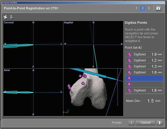
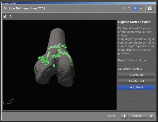
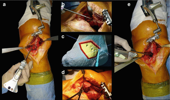
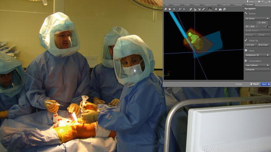
Once all plan is set, at the operation room, the navigation computer is best positioned opposite the surgeon approximately 5 feet away from the patient. The camera is located over the patient and directed downward at 45°. After surgical exposure, two 3 mm Apex pins are placed on the affected bone at least 3 cm away to the osteotomy programmed line in an area not affected for the tumor based in the preoperative planning. A navigation tracker is fixed to the pins. An image-to-patient registration to match precisely the operative anatomy and preoperative virtual CT images is performed by paired points (Fig. 8.2) and surface points (Fig. 8.3). In terms of surgical steps, 4 or at least 3 landmark points are identified in the affected bone based in the surgical exposure and anatomic visible points. Then, surface mapping of the bone is performed to reduce any mismatch between preoperative images and the patient’s anatomy, using at least 80 surface registration points in unaffected bone. After registration, the surgeon must double check with the navigation pointer if the surface of the patient`s bone in real time correlates on the virtual preoperative images. Afterwards, using the navigation pointer the osteotomies are marked in the surface of the bone (Figs. 8.4 and 8.5). The directions of the bone cuts are determined with the pointer and the osteotomies are performed with an oscillating saw or an osteotome.


Fig. 8.2
Point to point registration

Fig. 8.3
Surface refinement registration

Fig. 8.4
(a–c) Pointer marking multiplanar osteotomy, (d) Bone tumor resection, (e) Bone defect after resection

Fig. 8.5
Bone tumor navigation
Finally, another advantage that has this procedure, is the reconstruction of the bone defect, using computerized assistance. This is mainly accomplished when the reconstruction is done with bone allografts. The procedure is to reproduce the same planned cuts used during tumor resection, about the Allograft. This way can the defect can be reconstructed with more precision than the conventional way (allograft tailored by hand without any guidance), as if they were pieces of a puzzle.
Results
For this review, we analyzed our first 78 cases performed during the first 2 years of the utilization of this technology. All patients were followed for at least 2 years after surgery.
In four of the 78 patients (5 %) the navigation was not carried out due to technical problems (1 pelvis, 1 humerus and 2 femurs). In two cases the crash was secondary to software problems. In the remaining two cases the crash was secondary to hardware problems. One of the software technical crash happens in a case was the computer does not recognize one letter of the patients last name (the spanish ñ). The remaining software crash was that we tried to navigate the position of the osteosynthesis plate for the reconstruction, and the information to perform this exceeds the capacity of the computer navigation system. The hardware failures were related to broken trackers undetected during the procedure. All failures occurred during the first year of the utilization of this technology.
The mean time for navigation procedures during surgery was 31 min (range 11–61). However, time was improved during the learning curve. Therefore, the latest computer assisted surgeries, were faster than the first ones. We observe than in pelvic tumors the mean time for navigation procedure as 41 min (range 23–61), but the mean time for navigation procedure in extremities tumors was 27 min (range 11–54).
Of the 78 cases where the navigation was performed, the median registration error was 0.6 mm (range 0.3–1.1). This range of registration error was constant during time, from the first navigation through the last one.
Stay updated, free articles. Join our Telegram channel

Full access? Get Clinical Tree


