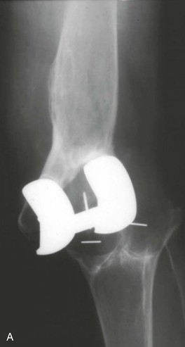
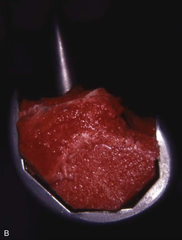
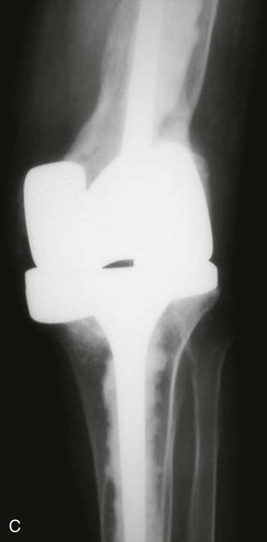
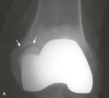
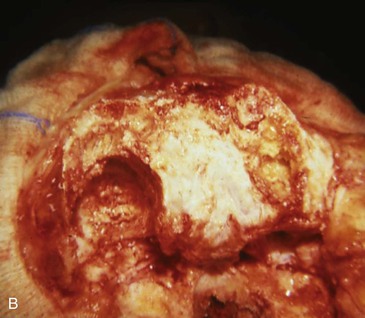
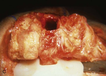
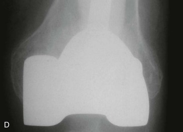
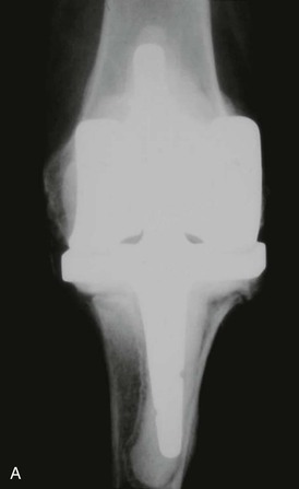
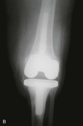
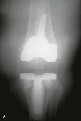
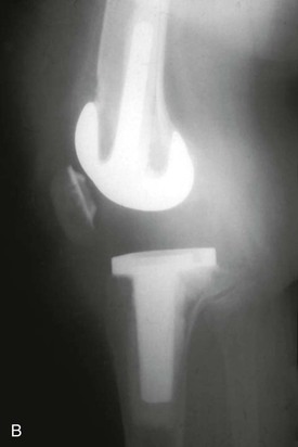
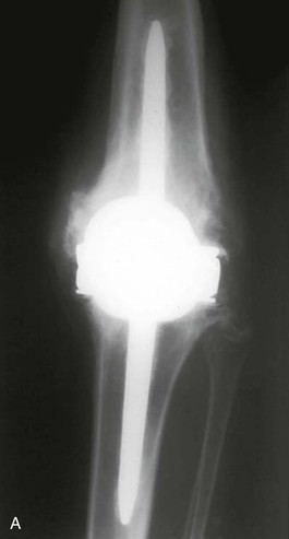
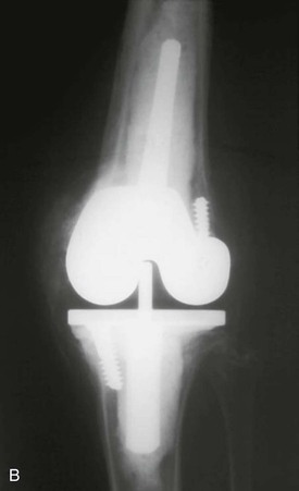
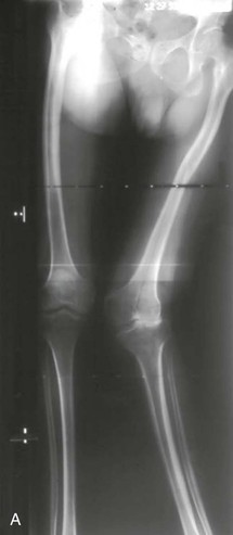
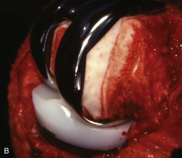
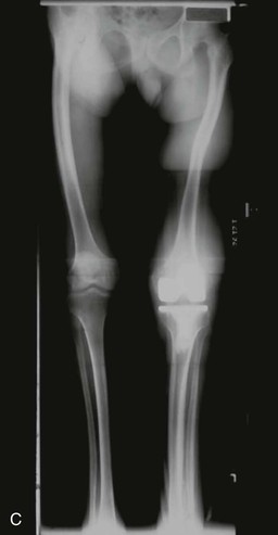
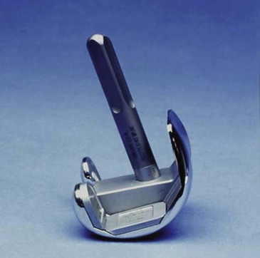
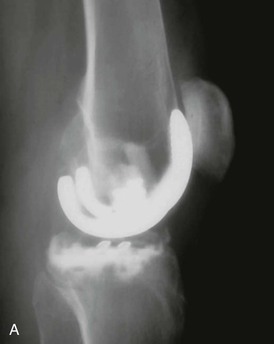
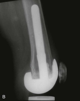
Options available for reconstitution of deficient bone stock on the femoral side include bone grafting, cement alone, cement plus screw augmentation, augmented components, and custom components. Bone grafting is appropriate for all contained defects. The graft can be in the form of morsels, solid blocks, or a combination of both. Early in my experience with patients with rheumatoid arthritis, I discovered that if juxtaarticular cysts were filled with cement, they could eventually develop an enlarging zone of demarcation at this interface and component loosening could occur (Figure 11-1). Histologic examination of the bone–cement interface in tissue retrieved at revision showed that the rheumatoid process had recurred at this interface, contributing to the loosening process (see Chapter 10). This finding convinced me that all juxtaarticular cysts, in both rheumatoid and osteoarthritic disease, should be curetted free of fibrous tissue and grafted with cancellous bone, which usually can be obtained from the standard bone resections. Follow-up radiographs show good incorporation of bone graft in these situations. In the presence of uncontained defects, the use of long stems and cement alone sometimes can suffice to achieve adequate prosthetic fixation (Figure 11-2). Care must be taken with this technique, however, to not distort the joint line. The most common error is elevation of the joint line combined with the use of thick tibial inserts to restore extension stability. Joint line elevation leads to patella baja and disturbance of the kinematics of the collateral ligaments (Figure 11-3). It is important to use a method that restores the femoral joint line to its more normal location. A simple way to accomplish this has been used for many years—the incorporation of bone screws into the cement mantle. Either cortical or cancellous screws are used, depending on the quality of the distal bone. The screws are allowed to protrude to the level at which they support the trial femoral component in its proper distal and varus-valgus position (Figure 11-4). This position can be estimated by determining the level of the normal joint line based on the opposite knee and measured as the distance from a fixed bony landmark such as the lateral epicondyle. The distance is usually 2 to 3 cm distal to the epicondyle, depending on the patient’s size. The proper level also can be assessed by its effect on the proximal distal positioning of the patella relative to the joint line. Valgus knees with a deficient lateral condyle often present a problem in restoration of the joint line in a primary situation. Classically, the severe valgus knee has a deficient lateral condyle (see Chapter 5). This hypoplasia accounts for much of the valgus deformity. The surgeon must avoid resecting back to the level of the condylar deficiency because it will result in elevation of the joint line. Resection should be based on the normal medial side, resulting in the need for lateral augmentation (see Figures 5-8 and 5-9). An effective and efficient way to accomplish this is with one or two cortical screws inserted into the sclerotic bone of the distal condyle. Properly placed screws support the trial femoral component in its proper distal and valgus positioning and perform the same function when the real component is cemented (Figure 11-5). I have used this method for more than 40 years without adverse effects. Concern is often raised about the use of dissimilar metals with the screw and femoral component. This concern is obviated if the screw is titanium. I believe, however, that chrome-cobalt or stainless steel screws can be used safely in conjunction with a chrome-cobalt femoral component because the encompassing bone cement protects the complex from potential adverse effects of dissimilar metals in close proximity. Thomas Thornhill and I introduced femoral augmentation wedges with the Omnifit revision total knee system in the late 1980s. Modular wedges are now available with virtually all revision systems (Figure 11-6). These can be affixed to either distal or posterior femoral condyle in various sizes. In some systems, the wedges are mechanically fixed to the femoral component, and in others, they are cemented. Both methods appear to be effective (Figure 11-7). Depending on the system, distal wedges as thick as 20 mm and posterior wedges as thick as 12 mm are available. Large-bulk femoral allografts are necessary for restoration of bone deficiency in extreme cases. My preferred method for their use is to affix the allograft to the host femur via the intramedullary alignment system and proceed with the bone cuts on the allograft in situ. Alternatively, the surgeon could prepare the allograft for the femoral component on a back table and subsequently couple it to the host bone via the long stem of the femoral component. The most difficult part of this technique is establishing proper femoral rotation at the host allograft junction and restoring both leg length and the position of the joint line relative to the quadriceps mechanism. I prefer to use a press-fit long stem if it can achieve excellent initial fixation. Otherwise, cemented stems are appropriate (Figure 11-8) and are also used for most smaller bulk grafts.
Bone Stock Deficiency in Total Knee Arthroplasty
Femoral Deficiency
Bone Grafting
Cement Alone
Cement Plus Screw Augmentation
Augmented Components
< div class='tao-gold-member'>
Related posts:
 Flexion Contracture Associated with Total Knee Arthroplasty
 Total Knee Arthroplasty in Rheumatoid Arthritis
 Sepsis and Total Knee Arthroplasty
 Patellofemoral Complications Associated with Total Knee Arthroplasty
 Staying Out and Getting Out of Trouble During Total Knee Arthroplasty
 Posterior Cruciate Ligament Retention Versus Substitution
![]()
Stay updated, free articles. Join our Telegram channel

Full access? Get Clinical Tree


Bone Stock Deficiency in Total Knee Arthroplasty
Chapter 11
Only gold members can continue reading. Log In or Register to continue
