19 Bone disorders
Cases relevant to this chapter
37, 41, 45, 53, 63, 73, 82, 92, 95, 98
Essential facts
1. Osteoporosis is the most common metabolic bone disorder.
2. It is important to look for underlying causes of osteoporosis.
3. Osteomalacia is under-diagnosed in the UK.
4. The commonest cause of osteomalacia is vitamin D deficiency.
5. Paget’s disease is due to overactivity of the osteoclast.
6. Osteonecrosis typically presents with pain and causes morbidity in young active people.
7. Causes of osteonecrosis include increased intra-osseous pressure, fat emboli, external compression of blood supply, direct osteocyte death, mechanical stress and increased thrombotic tendency.
8. Magnetic resonance imaging is the most sensitive investigation for early osteonecrosis.
Osteoporosis
Osteoporosis is the most common bone disorder and the pathophysiology is discussed in detail in Chapter 2. It is a significant cause of morbidity, increased disability and mortality, and imposes a major economic burden on the NHS. There is a 10–30% increase in mortality in the 12 months following a hip fracture. Osteoporosis is defined as a condition of skeletal fragility characterized by reduced bone mass and microarchitectural deterioration predisposing a person to an increased risk of fracture. The following mechanisms are responsible either alone or in combination: a failure to achieve adequate peak bone mass, an increase in bone resorption and a reduction in bone formation. It is more common in women and in the Caucasian population. Post-menopausal bone loss is the most significant cause of osteoporosis (Box 19.1).
Clinical features
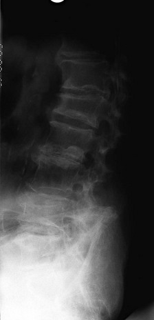
Osteoporosis is asymptomatic. Fragility fractures are the main consequence of osteoporosis, and can present either with fracture or with pain or loss of height with development of a thoracic kyphosis (Fig. 19.1). If the osteoporosis is secondary to another disorder, for example Cushing syndrome, features of the underlying disease may be the initial presenting complaint. The bones most frequently affected by a fragility fracture are the hip, vertebra and wrist. The risk is related directly to age, and is due to a combination of age-related bone loss and an increased rate of falls (Box 19.2).
Box 19.2
Risk factors for the development of osteoporosis
Aetiology
As described in Chapter 2, bone remodelling is a dynamic process, and the resorption and laying down of new bone are tightly coupled processes. In osteoporosis there is an imbalance of osteoclast and osteoblast activity. There may also be an increase in the initiation of new bone remodelling cycles (activation frequency). The resorption phase is faster than the formation phase, which can further contribute to osteoporosis when the activation frequency is high. Genetic factors contribute strongly to peak bone mass and to the rate of bone loss after peak mass has been achieved. Oestrogen has a central role in both men and women, but men do not have the same dramatic changes in sex hormone levels during middle age as women do (Table 19.1).
Table 19.1 Underlying mechanisms of osteoporosis
| Disease | Mechanism |
|---|---|
| Post-menopausal osteoporosis | Increased rate of remodelling, uncoupling of bone formation and resorption, increased osteocyte apoptosis, low oestrogen increases T-cell production of IL-1 and TNFα (both are osteoclastogenic), low oestrogen reduces osteoprotegerin (a regulator of bone turnover) |
| Hyperparathyroidism | Increased bone turnover |
| Hyperthyroidism | Increased bone turnover |
| Cushing’s disease | Uncoupling of bone resorption and formation |
| Corticosteroid treatment | Uncoupling of bone resorption and formation; increased osteocyte apoptosis, renal calcium loss and secondary hyperparathyroidism |
| Vitamin D deficiency | Direct and secondary hyperparathyroidism |
| Calcium deficiency | Secondary hyperparathyroidism |
Investigations
Box 19.3 outlines the investigations for osteoporosis.
Box 19.3
Investigations for osteoporosis
• Bone profile (calcium, phosphate, alkaline phosphatase, parathyroid hormone)
• Multiple myeloma screen (erythrocyte sedimentation rate, serum immunoglobulins and protein electrophoresis, urinary Bence Jones protein)
• Consider thyroid function tests, coeliac screen, urinary cortisol, testosterone, oestradiol, luteinizing hormone, follicle stimulating hormone, prolactin
Bone turnover markers
Bone turnover marker levels (Table 19.2) are affected by a number of factors:
• Significant inter- and intra-individual variation
• Turnover parallels growth velocity during childhood and fracture healing
• Seasonal variation (follows vitamin D levels through seasons)
• Medication (bisphosphonates, corticosteroids)
• The presence of other disease, overt or subclinical, that affects bone turnover (Paget’s disease, thyroid disease, hyperparathyroidism, osteomalacia)
• Renal failure causes a false increase in bone turnover markers.
Table 19.2 Bone turnover markers
| Resorption | Serum C– and N-telopeptides of type I collagen crosslinks (urine) |
| Formation | Pro-collagen type I peptide (PINP), bone-specific alkaline phosphatase, osteocalcin, C– and N-terminal pro-peptides of type I collagen |
Treatment
The aim of treatment is to reduce the incidence of fragility fractures. A readily available on-line calculator can be used to establish absolute risk of future fracture (FRAX™), which is an improvement over the T score, but there are still limitations. Lifestyle modification measures should be discussed with all patients, including improving calcium intake (equivalent of 1 pint of milk a day or 600 mg/day), supplemental vitamin D (50–75 nmol/l), weight-bearing exercise, healthy body mass index, smoking cessation and reduction of alcohol consumption if excessive. Balance and exercise classes promote increased bone density, but also help in fall prevention. Hip protectors have been advocated, but compliance is poor and effectiveness has been questioned. Current drug options are shown in Table 19.3, but fracture still occurs despite appropriate treatment.
Table 19.3 Drugs for osteoporosis, with mechanism of action and side effects
| Drug | Mechanism of Action | Side Effects |
|---|---|---|
| Calcium and vitamin D | Reduces hyperparathyroidism of increasing age | Hypercalcaemia |
| Bisphosphonates | Inhibition of osteoclast activity, increased osteoclast programmed cell death, slowing of the remodelling cycle allowing full mineralization of new bone to occur | Oesophagitis |
| PTH (teriparatide) – synthetic N-terminal portion of PTH | Large increase in bone formation (anabolic) and a modest increase in resorption; increased periosteal apposition (deposition of bone on the surface to increase strength) | Headaches, rarely; hypercalcaemia, nausea, leg cramps |
| Strontium ranelate | Mechanism is not fully understood. Has anti-resorptive and anabolic effects | Diarrhoea, headache, nausea, deep vein thrombosis (DVT) |
| HRT (hormone replacement therapy) | Reduces bone resorption by blocking cytokine signalling to the osteoclast | Risk of breast cancer, DVT, increased cardiovascular risk |
| SERMs (selective (o)estrogen receptor modulators) | Reduces bone resorption | Increased DVT risk and hot flushes (reduction in breast cancer risk) |
| Denosumab | Inactivates RANKL and so inhibits osteoclast maturation | Usually well tolerated, but may cause diarrhoea, headache, nausea and tiredness. |
Stay updated, free articles. Join our Telegram channel

Full access? Get Clinical Tree


