, Paul D. Siney1 and Patricia A. Fleming1
(1)
The John Charnley Research Institute Wrightington Hospital, Wigan, Lancashire, UK
Demarcation on the radiographs is seen as a dark line between the radiopaque cement and the bone of the acetabulum. … the width was measured on the radiographs … and its distribution categorised into three zones I, II, or III. The radiographs were also scrutinised for bulk migration.
The complexity of this aspect of total hip arthroplasty was clearly documented by DeLee and Charnley [1]. A number of factors, either singly or a combination, affect the result. Design, materials, surgical technique, the method of component fixation, patient’s activity level, the length of follow-up, wear and mechanical changes resulting, as well as tissue reaction to wear particles, radiographic appearances, clinical results, findings at revision surgery and the histology of the bone-cement interface, are some of the important issues.
Demarcation of the Cup
Radiographic appearances are best recorded using accepted terminology without any attempt to correlate them to, or combine them with, the clinical results. The DeLee – Charnley description and classification [1] is accepted as the standard. The hemisphere of the cup is divided into three segments: I = 45°: II = 90°: III = 45° (Fig. 28.1). Why this unequal division is by no means clear.
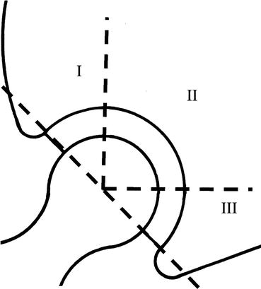
Fig. 28.1
Cup demarcation grading of DeLee and Charnley, showing division of hemisphere of acetabular bone-cement interface into zones, type I, II, III (Reproduced with permission from DeLee and Charnley [1])
The description is clearly one of “demarcation … seen as a dark line between the radiopaque cement and the bone of the acetabulum.” Histological examination revealed a fibrous tissue layer between bone and cement. This layer varied in appearance. “Thick layers of soft fibrous tissue …frequently alternate with thin layers of dense fibrous tissue … often the cement makes load–bearing contact with the underlying bone which closely resembles fibrocartilage.
The conclusion was:
“The vast majority with demarcation were symptomless.”
“… where demarcation was associated with migration … technical explanation or low grade sepsis was responsible.”
“better surgical technique may increase the number of cases showing no demarcation.”
Correlation Between Radiographic Appearances and Operative Findings [2]
This study was undertaken to establish the correlation between the radiographic appearances and the operative findings at the bone-cement interface of the cup of the Charnley LFA.
Method of Testing for Cup Loosening
The margin of the cup and the bone cement junction is exposed using Charnley gouges: This may include removal of the marginal osteophytes. The acetabulum retractor introduced into the margin of the obdurator foramen exposes the inferior margin of the cup where wear debris usually accumulates. The socket tester is screwed into one of the cup holder locating holes. Gentle rotating movement is applied to the tester while the bone-cement junction is observed. Movement, and at times fluid ejection, at the bone-cement interface is taken as evidence of cup loosening. (Extensive demarcation of the outer one third of the cup and gross wear allowing elastic deflection of the UHMWPE cup wall will produce a similar effect.) More than one area of the bone-cement junction must be examined.
Related posts:

Stay updated, free articles. Join our Telegram channel

Full access? Get Clinical Tree


