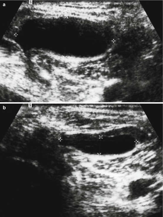
Fig. 11.1
Example of a Baker’s cyst with a possible ball valve mechanism due to large quantities of fibrin within the cyst as assumed by Jayson and Dixon [8]
In an anatomical study, Rauschning [3] found a communication between the knee joint and Baker’s cysts in 58 of 108 knees. The communication becomes progressively wider with increasing flexion as the gastrocnemius tendon was pulled away from the femoral condyle. On extension the capsule of the knee joint was compressed between the tendon and the femoral condyle. This result can explain the decreasing pressure in the Baker’s cysts under knee flexion. Because of the anatomical structure of the communication between the cyst and knee, Rauschning contradicted the hypothesis of a valve mechanism of the Bunsen type. Considering the observation of Pindler [10] who could empty all 14 popliteal cysts into the joint cavity during anterior synovectomy in spite of containing fibrin masses, Rauschning also contradicted the hypothesis of a true valve mechanism of the ball type. Instead, he interpreted the closing mechanism as a functional action concerning both directions and excluded a unidirectional valve mechanism.
Some other study results are also in contradiction to a unidirectional valve mechanism. Wilson et al. [11] found that air, injected in a popliteal cyst, promptly passed into the joint even if it is not possible to empty the cyst into the joint. Maudsley and Arden [12] described an activity trespassing into the knee joint after injection of yttrium into a popliteal cyst. In ten patients Smith et al. [13] measured activity distribution into Baker’s cysts after RSO of the knee using single-photon emission computed tomography (SPECT) and found 0–40 % of the activity in the cysts. Up to 46 h, repeated measurements over time showed no activity accumulation; during this time the patients were confined to bed.
11.3 Treatment Results of Baker’s Cysts After RSO
Up to now, only a small number of studies described the effect of RSO of the knee joint to Baker’s cysts. Grahame et al. [14] treated 15 patients: 13 showed a Baker’s cyst, and in 2 cases the cyst extended into the calf. In 4 of the 13 patients, the cyst disappeared. The time difference of control and treatment was between 6 and 11 months.
Topp et al. [15] described a study of 112 RSO of knee joints; the treatment results were controlled up to 5 years. Twelve patients had a popliteal cyst or synovial rupture or both, and in all cases the lesion disappeared or decreased in size.
In a prospective study of 150 RSO of knee joints with Baker’s cyst, Mödder [16] reported on 87 disappeared cysts after the first RSO. Another 57 cysts disappeared after a second RSO. Time difference of control and treatment was not mentioned.
Our own data of 60 RSO with Baker’s cysts in 45 patients showed a significant reduction of the median cyst volume of 73 % after a median follow-up of 6 months (Fig. 11.2). In 25 % of cases the cyst disappeared, and in 55 % the volume was reduced, whereas in 20 % the volume increased [17].


Fig. 11.2
Volume reduction of a Baker’s cyst due to RSO of the knee joint. (a) Baker’s cyst before RSO. (b) Baker’s cyst 5 months after RSO
11.4 Risk Estimation for Baker’s Cyst Rupture
Neither of the above-cited treatment studies reported any complication corresponding to Baker’s cyst. Nevertheless, Asavatanabodee et al. [18] described a Baker’s cyst rupture in 5 of 133 treated knee joints; time difference between RSO and rupture was not mentioned. In addition, Davis and Jayson [19] reported two cases of a knee joint rupture occurring 43 and 63 days after RSO.
The most severe complication would be the rupture of a Baker’s cyst directly after RSO, draining the activity into the calf. Therefore, for risk estimation not only the probability of cyst rupture but also the quantity of activity present within the fluid inside the cyst needs to be assessed. Standard of care mandates the immobilization of the knee joint for 48 h after RSO, so cyst rupture based on external stress can be expected earliest after the end of this immobilization period.
Because of aggravation of symptoms and recurrence of effusion, a puncture of the knee joint in one patient and of the Baker’s cyst in another patient had to be performed in our institution to exclude infection 12 days after RSO with the possibility of activity measurement in the aspirated fluid. Table 11.1 depicts the results of activity per ml aspirated fluid, decay-corrected activity per ml 48 h after RSO of the aspired fluid, and activity in the whole lumen of the knee joint and Baker’s cyst due to the injected activity 48 h after RSO. In addition, for risk estimation, the activity per 100 ml fluid 48 h after RSO as a worst case was calculated due to the activity in the aspirated fluid.
Table 11.1
Activities in knee joint fluid and Baker’s cyst fluid, respectively, 12 days after RSO and decay-corrected activities 48 h after RSO
Aspirated activity/ml [kBq] | Decay-corrected aspirated activity/ml 48 h after RSO [kBq] | Activity/100 ml 48 h after RSO [kBq] | Injected activity 48 h after RSO [kBq] | |
|---|---|---|---|---|
Patient 1 (joint puncture) | 3 | 40 | 4,000 | 137,000 |
Patient 2 (cyst puncture) | <1 | <13 | <1,300 | 120,000 |
The results show a huge difference between the calculated activity inside the joint due to the injected activity and the measured activity of the aspirated fluid with decay correction. This difference can be explained by the fast phagocytosis of the injected radiocolloids into the synovial membrane. Therefore, also in the unlikely case of a rupture of the Baker’s cyst, the emptied activity into the calf is very low, and a distinctive damage of the popliteal and calf tissue should not be expected.
11.5 Discussion
Initially Baker’s cyst was classified as an absolute contraindication for RSO of the knee joint because of some reports of spontaneous rupture, especially corresponding to patients with recurrent joint effusion [20–22]. Even today some authors still classify Baker’s cyst as an absolute contraindication [23]. Nevertheless, 20 years ago, Mödder [16] introduced a more differentiated view of the topic. He suggested the use of ultrasound to differentiate between Baker’s cyst with and without a valve mechanism and classified only Baker’s cysts with valve mechanisms as an absolute contraindication.
Stay updated, free articles. Join our Telegram channel

Full access? Get Clinical Tree


