12.1.6 Kyphosis and Back Extensor Strength
Spinal column is supported and moved with four major muscle groups including extensors, flexors, lateral flexors, and rotators [28]. Back extensor muscles are the main supportive muscles, which extend the spine and maintain the posture. Back extensor strength has been considered highly important in patients with osteoporosis. Osteoporotic women have been found to have significantly lower back extensor strength than healthy women [29], and back extensor strength demonstrated a negative correlation with kyphosis [30, 31].
Sinaki et al. evaluated back extensor strength, bone mineral density (BMD), and physical activity scores in a cross-sectional study involving 65 women [30]. Back extensor strength had a significant negative correlation with thoracic kyphosis and positive correlations with lumbar lordosis and sacral inclination. However, BMD and physical activity scores did not show any significant correlations with the radiographic factors. These results indicated that the stronger the back extensors, the smaller the thoracic kyphosis and the larger the lumbar lordosis and sacral inclination. In another cross-sectional study by Mika et al., 189 female subjects were grouped by their BMD, and various parameters were compared between the groups [31]. Multivariate analyses of back extensor strength and BMD showed that only back extensor strength affected thoracic kyphosis. There was no correlation between back extensor strength and BMD. A significant difference in back extensor strength was observed only between the osteoporotic and osteopenic groups. They concluded that the severity of thoracic kyphosis might be especially affected by changes in back extensor strength.
Besides these articles that demonstrated significant correlations between back extensor strength and thoracic kyphosis, we have demonstrated associations between lumbar lordosis and back extensor strength [32]. We assessed the association of osteoporotic spinal deformities with back strength in a total of 206 elderly women in Akita, Japan, and in Minnesota, USA. We found that thoracic kyphosis and lumbar lordosis were higher in the Minnesota group than in the Akita group. In the Akita group, multiple regression analysis showed that the angle of lumbar lordosis correlated significantly with back extensor strength, indicating the potential importance of strengthening the back extensors for improving or maintaining lumbar lordosis. The differences identified in the shape of sagittal spinal curvature from two geographic areas of the world might be due to the difference in race and lifestyles.
Based on these findings from several studies, back extensor strength is an important determinant of posture, and preparing strong, natural extrinsic support for the spine seems to be important to decrease the incidence of spinal deformity.
12.1.7 Measurement of Back Extensor Strength
Appropriate measurement of trunk muscle strength is required for the correct evaluation of the performance of the spine. Measurement of muscle strength is roughly classified into isometric and isokinetic procedures. Isokinetic measurement of muscle strength can evaluate physiologic muscle contracture with spinal motion, but the measurements were demonstrated to vary depending on the posture, and the apparatus is relatively expensive. On the other hand, measurement of isometric muscle strength provides reliable and accurate results with the appropriate device, and the device is relatively simple and easy to move. Smith evaluated the repeatability of the methods comparing the isometric and isokinetic strengths of trunk extensors, flexors, and rotators and concluded that the strength data were reliable, and there was no significant difference between the procedures [33].
Measurements of back extensor strength and back-strengthening exercises are better if specific instruments are used to measure trunk strength in sitting positions with various spinal postures [34]. However, the authors’ techniques used for measurement of back extensor strength and procedures for back-strengthening exercises in a prone position have been previously established as safe and reliable. During measurement and exercise, a prone position allows back extensors to work without contributions from the musculature of the lower extremities.
The authors use a custom-made strain gauge type back isometric dynamometer. The concept was originally described by Limburg et al. [35]. Subjects lie prone with the hips and knees in the neutral position and the arms fully extended at the elbow. No immobilizing straps are necessary with proper placement of the padded transducer head. Its upper edge is aligned with the superior borders of the scapulae across the midline. During the measurements, subjects raise their hands from the table to prevent the unwanted contribution of upper extremity musculature to the upward force generated by the trunk against the transducer head. Direct contract between the pelvis and the table must be maintained throughout the measurements. The extensor muscles are contracted for 5 s, with no initial upward jerking motions directed at the transducer. The maximum force generated during exertion is processed by the measuring device. The patient is allowed one warm-up trial, which is followed by three successive maximal effort trials separated by 1-min resting periods. The maximal force achieved is then recorded (Fig. 12.2). A more convenient and easy method for evaluating isometric back extensor strength in daily clinical practice has also been reported. A handheld dynamometer fixed with a tripod has been reported to be reliable for the assessment of back extensor strength in women with osteoporosis and vertebral fractures [36].
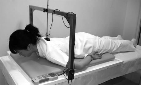
Fig. 12.2
Measurement of isometric back extensor strength with custom-made apparatus with dynamometer
12.2 Effects of Back-Strengthening Exercise
12.2.1 Bone Mineral Density
Back extensor strength demonstrated a positive correlation with BMD of the spine [37–39]. Iki et al. reported that greater trunk extensor torque reduced bone loss independently of age, body size, and vitamin D receptor genotype [37]. Zhou showed a correlation between muscle strength and BMD using isometric and isokinetic testing modes, demonstrating an age-dependent reduction in BMD and muscle strength throughout early menopause [40]. Sinaki et al. conducted a randomized, controlled trial evaluating the effect of a less demanding exercise program for back extensor muscles using a backpack on muscle strength and bone mineral density [41]. They found that postmenopausal bone loss was unaffected by a modest exercise program despite an increase in muscle strength and concluded that back muscle exercises might be ineffective in retarding vertebral bone loss in ambulatory, healthy, postmenopausal women.
12.2.2 Posture
Several studies have demonstrated the effect of exercise on posture [42–51]. Itoi and Sinaki conducted a study evaluating the effect of a back-strengthening exercise with a weighted backpack for 2 years on spinal curvature [42]. They randomly assigned 60 subjects to either an exercise or a control group and found that back extensor strength increased significantly in both the exercise and the control groups, but no radiographic measurements were significantly different between these groups. The significant increase in back extensor strength in both groups of healthy women suggested that the original grouping did not accurately reflect the amount of exercise. Thus, the 60 subjects were reclassified for comparison on the basis of increase in back extensor strength: 27 with more than or equal to the mean increase of 21.1 kg and 33 with less than 21.1 kg. Furthermore, each of these groups of subjects was subdivided on the basis of degree of thoracic kyphosis. Among the subjects with substantial thoracic kyphosis, those with a significant increase in back extensor strength had a significant decrease in thoracic kyphosis, whereas those with a small increase in strength had a nonsignificant increase in thoracic kyphosis. The authors concluded that increasing the back extensor strength in healthy estrogen-deficient women helps decrease thoracic kyphosis.
Pilates exercise is a “mind-body exercise” that has been used since early in the twentieth century. It focuses on improving strength, core stability, flexibility, muscle control, posture, and breathing [52]. Kuo et al. assessed the changes in sagittal spinal posture in 34 older volunteers after a Pilates-based exercise program for 10 weeks [45]. They found that immediately after the exercise program, participants stood with slightly decreased thoracic flexion and sat with slightly increased lumbar extension. The individually designed Pilates-based exercise program was feasible for healthy older adults, and the high attendance rate supports the suitability of the exercise program over a long period.
Greendale et al. conducted a randomized, controlled trial involving 118 participants with a kyphosis angle of at least 40° and assessed whether a specifically designed yoga intervention could reduce hyperkyphosis [53]. The active treatment group attended hour-long yoga classes 3 days per week for 24 weeks. At the final follow-up, participants randomized to yoga experienced a 4.4 % improvement in kyphosis angle. Based on the results of this study, the decrease in the flexicurve kyphosis angle in the yoga treatment group showed that hyperkyphosis is remediable, a critical first step in the pathway to treating or preventing this condition. However, Sinaki reported three healthy persons with low bone mass who developed new pain and fractures after participation in yoga flexion exercises and cautioned that flexion yoga positions in patients with osteopenia or osteoporosis could lead to an increased risk of vertebral fractures [54].
Benedetti et al. performed a clinical study to systematically compare the effects of a physical activity program that specifically addressed the flexed posture with a nonspecific exercise program for 3 months and concluded that the program significantly improved postural alignment and musculoskeletal impairment of the elderly [47].
Ball et al. evaluated the progression of kyphosis with age and found that kyphosis increased with age in healthy women, with the greatest difference observed between women 50 and 59 years of age. They then conducted a 1-year prospective, descriptive analysis of the effect of extension exercises on posture in women 50–59 years of age. The progression of kyphosis was greater in women who did not perform extension exercises than in those who performed extension exercises three times per week for 1 year [46].
We have conducted a randomized study assessing the effect of back extensor strength on spinal sagittal curvature [51]. Lumbar lordosis at the neutral position significantly increased in the exercise group. The increase in lumbar lordosis was also significantly larger compared with the control group (Fig. 12.3). However, no significant change was found in thoracic kyphosis. In this study, back-strengthening exercise was shown to be effective in improving spinal deformity by increasing lumbar lordosis.
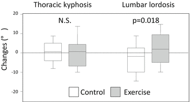
Fig. 12.3
Changes in thoracic kyphosis and lumbar lordosis after back exercise intervention for 4 months (Modified from Hongo et al. [51] with permission)
12.2.3 Vertebral Fractures
A cross-sectional study of patients with osteoporosis showed a negative association between back extensor strength and both kyphosis and the number of vertebral fractures, suggesting that increasing back strength could reduce the risk of vertebral fracture for the osteoporotic spine [55].
As described above, Itoi et al. evaluated the effect of back-strengthening exercise on improvement of kyphosis and found that kyphosis improved with the exercise at the 2-year follow-up [42]. The authors then investigated the long-term protective effect of strengthening the back muscles on vertebral fractures. Subjects performed progressive, resistive back-strengthening exercises for 2 years and were followed up at 2 years and 10 years. The authors found that the back extensor strength in the exercise group was still significantly higher even 8 years after cessation (Fig.12.4a) and the relative risk of compression fracture was 2.7 times higher in the control group than in the exercise group (Fig. 12.4b). They concluded that stronger back extensors achieved through exercises reduced the risk of vertebral compression fractures (Fig. 12.4a, b) [56].
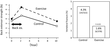
Fig. 12.4
(a) Change in back extensor strength at 10-year follow-up after cessation of exercise for 2 years. (b) Incident of vertebral fractures at 10-year follow-up (Modified from Sinaki et al. [56] with permission)
Another study also showed the effect of back-strengthening exercise on the reduction of re-fracture after percutaneous vertebroplasty. In this retrospective analysis, the authors reviewed the data of patients with osteoporosis treated by vertebroplasty. The results showed that an exercise program for osteoporosis including back strengthening after percutaneous vertebroplasty decreased fracture recurrence [57].
Sinaki and Mikkelsen conducted a study comparing treatment programs with extension exercises, flexion exercises, combined exercises, and no therapeutic exercises [58]. The study suggested that a significantly higher number of vertebral compression fractures occurred in patients with postmenopausal osteoporosis who followed a flexion exercise program compared with those using extension exercises. They concluded that extension or isometric exercises seem to be more appropriate for patients with osteoporosis.
12.2.4 Quality of Life
There have been multiple studies describing the effects of interventions on health-related QOL in older women with low bone mass [59–62]. Home-based exercises have been shown to improve QOL scores [61, 62]. Group treatment including trunk extension exercises also improved psychological status, which is considered part of QOL [59]. However, the participants were instructed to engage in the home-based or institutional-based exercise for 45–60 min per day.
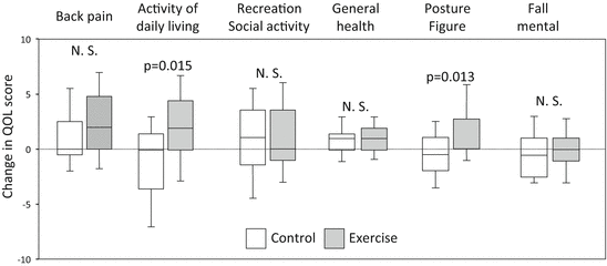
We conducted a randomized, controlled study in 80 postmenopausal women with osteoporosis to investigate the effect of a home-based, simple, low-intensity exercise. QOL scores increased significantly in the exercise group, while they remained unchanged in the control group. Significant improvements were observed in the scores for activities of daily living and posture in the exercise group compared with control group. Back pain also improved in both groups; however, there was no difference in the improvement of back pain between the groups (Fig. 12.5). Since physical activity was reported to correlate with back extensor strength, enhancing back extensor strength may have a direct effect on improvement of QOL [63]. In this study, the implementation of exercise was relatively favorable, with compliance of 73 % and no major adverse events; the exercise regimen can be safe and sustainable for elderly persons. However, this exercise may not be suitable for kyphotic patients who are unable to lie in a prone position.
Miyakoshi et al. then evaluated the effect of a simple and lower intensity back-strengthening exercise that can be performed by participants who are unable to lie in the prone position [64]. Two types of simple and low-intensity back extension exercises were performed by 31 postmenopausal elderly women. The authors found that 6 months of simple, low-intensity back extension exercises improved QOL and back extensor strength in patients with postmenopausal osteoporosis and vertebral fractures.
12.2.5 Optimum Intensity and Frequency of the Exercise
There have been some concerns related to the exercise prescription. Subjects in the previous studies were all healthy postmenopausal women, and the average weight used in these studies of healthy postmenopausal women was 22 kg, which was equivalent to 30 % of the maximum strength of the back extensors [41]. The same intensity of exercise is not recommended for patients with osteoporosis. As in any weight-lifting exercise program, the exercises need to be modified for osteoporotic patients to avoid pain and/or fracture. As long as it is effective in increasing the back extensor strength, a reduction in the intensity of exercise may be beneficial for patients with osteoporosis.
The effect of less resistance training on achieving significant improvements in trunk muscle strength is not well understood for elderly populations. Swezey et al. reported brief, progressively resisted, trunk isometric exercises with use of an inflatable vinyl ball for postmenopausal women with osteopenia and osteoporosis and found an improvement in trunk extensor strength [65]. Vincent et al. compared muscle strength after low-intensity (50 % of one repetition maximum (1RM)) and high-intensity lumbar exercises (80 % of 1RM) in 60- to 83-year-old adults [66]. It is interesting to note that the magnitude of the increases in lumbar extensor strength was greater with low-intensity lumbar exercise than with high-intensity lumbar exercise. These results suggest that lower intensity exercise provides a similar effect on muscular strength in elderly persons to high-intensity exercise. The authors demonstrated that exercise using a reduced weight was less effective in improving back extensor strength than exercise with an average weight of 14 kg, which was equivalent to 30 % of the maximum back extensor strength in a study with young women volunteers [67].
12.2.6 Author’s Recommendation: Low-Intensity Back-Strengthening Exercise
The low-intensity back-strengthening exercise regimen could be performed at home for a duration of approximately 5 min, and it has been demonstrated to be feasible, safe, and effective with high compliance for elderly individuals [63]. In the study, the exercise group with no additional weight had a significant increase in back extensor strength compared with the control group.
The back exercise regimen with low resistance should be considered in terms of feasibility, safety, and effectiveness for the elderly population with osteoporosis. In 1982, it was reported that the combination of a few exercises with avoidance of flexion can safely and effectively strengthen the fragile osteoporotic spine [68]. The author’s recommendation for the back exercise is just one exercise for back strength without performing the full program described in the initial study.
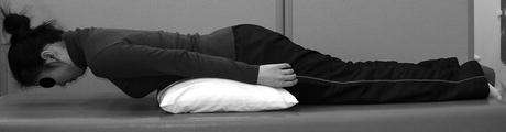
The selected single home-based exercise is done according to the procedure described previously [67–69]. Subjects are asked to lie in a prone position on a bed with a pillow under the abdomen such that the spine is slightly flexed. Following a warm-up exercise in which the spine is slowly extended with the aid of both arms ten times, the subjects are then asked to lift the upper trunk off the bed toward the neutral position for 5 s, with a 10-s interval between the contractions (Fig. 12.6). Each contraction is repeated 10 times as a set, with the duration of exercise ranging from 3 to 5 min. It is recommended that this exercise be performed as one set a day, 5 days a week. Back extensor strength and QOL, including physical activity, pain, and posture, can be expected to eventually improve [51, 63]. This effect was considered to be based on the weight of the head, upper extremity, and thoracic trunk.
 Vertebral Strength Changes as Assessed by Finite Element Analysis
 Spondylosis and Osteoporotic Vertebral Fractures
 Exercise and Fall Prevention
 Applications of Teriparatide for Fracture Repair and Osteosynthetic Surgery in Osteoporosis
 Collagen Cross-Links as a Determinant of Bone Quality
 Sarcopenia and Osteoporosis


Related posts:
Vertebral Strength Changes as Assessed by Finite Element Analysis
Spondylosis and Osteoporotic Vertebral Fractures
Exercise and Fall Prevention
Applications of Teriparatide for Fracture Repair and Osteosynthetic Surgery in Osteoporosis
Collagen Cross-Links as a Determinant of Bone Quality
Sarcopenia and Osteoporosis
Stay updated, free articles. Join our Telegram channel
Full access? Get Clinical Tree
