Fig. 5.1
Anatomy of the carotid artery
Frequency and Severity:
Contour of the bruit, especially in the <200 Hertz (Hz) frequency range, is poorly predictive of degree of stenosis [6, 7].
In the late 1960s, Carotid Phonoangiography was proposed as a means to predict degree of carotid stenosis (Fig. 5.2):
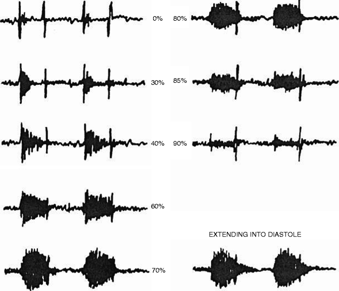
Fig. 5.2
Carotid phonoangiography in symptomatic patients, <200 Hz with projected degree of stenosis; X-axis time, Y-axis amplitude (Reproduced with kind permission from Springer Science + Busines Media from Lees [36])
Higher frequencies and bruit duration may improve the accuracy of carotid phonoangiography in more contemporary studies:
Frequencies greater than 340 Hz predict significant stenosis:
Duration greater than 200 ms predicted stenosis >60 %:
24/26 (92 %) patients with a duration >200 ms had peak carotid velocities >200 cm/s consistent with stenosis >60 % [11].
Differential diagnosis of carotid auscultory abnormalities [1]:
Venous hum:
A venous hum is a benign phenomenon resulting from venous vibration due to blood return along the jugular vein.
It is heard throughout the cardiac cycle, can be eliminated by firm pressure against the skin with the stethoscope, and is often abolished when the patient is in the supine position or turns their head to one side.
Most common in patients <20 years-old [12] or in patients with increased cardiac output, such as seen with anemia, thyrotoxicosis or pregnancy
Radiating valvular murmurs:
To differentiate carotid bruits from valvular murmurs, ausculatation of the precordium is required to listen for valvular murmurs such as aortic stenosis. The murmur of aortic stenosis, a harsh crescendo-decrescendo systolic murmur, will radiate from the right upper sternal border into the neck.
Carotid bruits do not radiate to the precordium.
Thyroid arterial bruit:
Auscultory sound related to increased blood flow to a hyperactive thyroid gland
Suspicion for this disorder should be high in patients with hyperthyroidism or an enlarged thyroid gland.
Murmurs resulting from stenosis of the great vessels in the thorax, such as aortic arch, brachiocephalic, the proximal left common carotid or left subclavian arteries, are typically best heard at the base of the neck over the ipsilateral subclavian artery. Stenosis of the subclavian artery may also result in a murmur that transmits to the axilla on the affected side.
It is important to note that an occluded carotid artery produces no bruit:
An occluded carotid artery may produce bruit in the opposite side through contralateral increased flow [5].
Obesity in today’s population creates difficulty in the carotid examination:
A continuous (systolic + diastolic) neck bruit may indicate ipsilateral high-grade stenosis or high grade stenosis or occlusion on contralateral side [9]:
A continuous bruit results from turbulence in both high flow systole and lower flow diastole.
In the case of contralateral carotid artery high-grade stenosis or occlusion, this is thought to occur through increased turbulence by the establishment of augmented collateral flow.
Auscultation for the presence of bruits is not a sensitive test for the presence of significant carotid atherosclerosis [16–21]:
Detection of a carotid bruit has a sensitivity of 36 % for stenosis of >60 % in asymptomatic patients in a multiethnic population [21].
Carotid bruit detection has a sensitivity of 24 % for stenosis of >50 % in asymptomatic patients referred to coronary artery bypass surgery [18].
Screening for carotid bruits has been reported with a sensitivity of 6 % for the presence of carotid artery plaque in asymptomatic patients [21].
Detection of a carotid bruit has good specificity [16–21]:
The specificity for ultrasound stenosis >60 % is 98 % in asymptomatic patients in a multiethnic population [18].
In another study, a carotid bruit had a specificity of 96 % for stenosis of >50 % in asymptomatic patients referred to coronary artery bypass surgery [18].
In a study of asymptomatic patients, detection of a carotid bruit was shown to have a 99 % specificity for the presence of atherosclerotic plaque on carotid ultrasonography [19].
However, it important to note that the detection of a carotid bruit does not predict side on which stroke will occur [22] or the degree of carotid lumen stenosis [16–19].
Carotid bruit detection has been shown to have good inter-observer reproducibility [23]:
Independent clinicians tend to agree whether or not a carotid bruit is present with a κ = 0.67.
Prevalence of Carotid Bruits
Carotid bruits have been reported in up to one-third of healthy young adults when using a stethoscope [24]:
These are usually false positives and are thought to be due to the relatively hyperdynamic young hearts.
Carotid Doppler
While two-dimensional images of the carotid artery are easily obtained and utilized to assess for the presence and location of carotid artery plaque, the degree of carotid artery stenosis is most accurately assessed using Doppler techniques to determine the velocity and pattern of carotid arterial flow:
Basic technique:
Accurately assess the velocity and pattern of flow
A high frequency 7–10 MHz linear array transducer
Mild stenosis:
Peak frequency below 4 kHz (120 cm/s), but some spectral broadening
Severe stenosis:
Peak frequency above 4 kHz and spectral broadening
Very severe stenosis:
High end diastolic frequency
See Fig. 5.4.
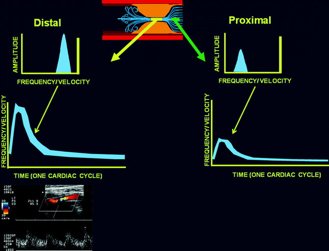
Fig. 5.4
Spectral waveforms of carotid Doppler signals proximal and distal to a stenosis showing both an increase in amplitude and frequency/velocity in the setting of a stenosis
Asymptomatic Versus Symptomatic Carotid Bruit
Carotid Bruits and Cardiovascular Prognosis
The National Cholesterol Education Program (NCEP) defines a coronary risk equivalent as any risk factor whose presence carries a 2 %/year risk or a 20 % over 10 year risk of a myocardial Infarction or cardiovascular death [27].
Recently, two meta-analyses have demonstrated the prognostic significance of asymptomatic carotid bruits [28, 29].
Experts have cited this work as highlighting the utility for neck auscultation to identify patients at high cardiovascular risk who may benefit from more intensive cardiovascular prevention strategies [30].
Carotid Bruits: Cardiovascular Morbidity and Mortality (Table 5.1):
Table 5.1
Carotid bruits and incident myocardial infarction and cardiovascular mortality
Risk ratio (95 % confidence interval [CI]) (carotid bruit vs. none)
With carotid bruit (95 % CI) (events/100 patient-years)
Without carotid bruit (95 % CI) (events/100 patient-years)
Myocardial infarction
2.15 (1.67–2.78)
3.69 (2.97–5.40)
1.86 (0.24–3.48)
Cardiovascular death
2.27 (1.49–3.49)
2.85 (2.16–3.54)
1.11 (0.45–1.76)
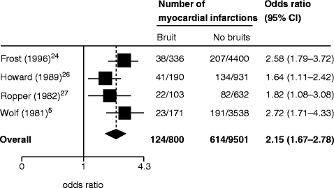
Presence of a carotid bruit in an asymptomatic individual portends a risk ratio of 2.15 for incident myocardial infarction (MI) [28]:
The rate of MI in patients with a carotid bruit is 3.69 events per 100 patient years (Fig. 5.5).
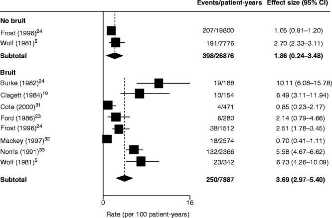
Fig. 5.5
Annual rate of myocardial infarction in asymptomatic patients with versus without carotid bruit (Reproduced with permission from Pickett et al. [28])
The rate of MI in Patients without Carotid Bruits is 1.86 events per 100 patient years (Fig. 5.5).
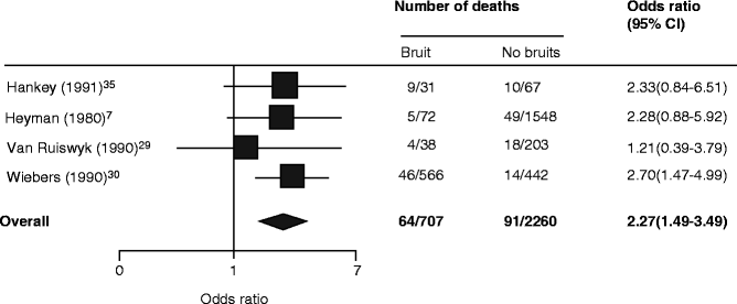
Presence of a carotid bruit portends a risk ratio of 2.27 of cardiovascular death [28]:
The rate of cardiovascular death in patients with a carotid bruit is 2.85 events per 100 patient-years (Fig. 5.7).
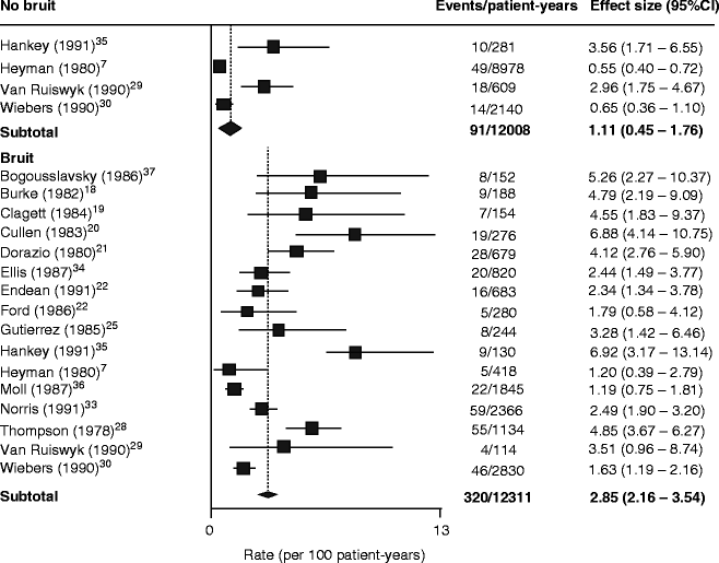
Fig. 5.7
Annual rate of cardiovascular death in patients with and without a carotid bruit (Reproduced with permission from Pickett et al. [28])
The rate of cardiovascular death in patients without a carotid bruit is 1.11 events per 100 patient-years (Fig. 5.7).
The presence of a carotid bruit meets the NCEP definition of a coronary artery disease risk equivalent (>2 %/year risk) with an annual rate of 2.85 myocardial infarctions per 100 patient-years.
Cerebrovascular Morbidity and Mortality (Table 5.2):
Table 5.2
Carotid bruits and risk of subsequent transient ischemic attack, stroke and death from strokeRelated posts:
 Insights into the Natural History of Atherosclerosis Progression
 Positron Emission Tomography: Imaging Inflammatory Atherosclerosis
 Factors Contributing to the Development of Atherosclerosis
 Intravascular Ultrasound
 Noncontrast Cardiac CT for the Detection of Calcified Atherosclerosis
 Coronary Atherosclerosis Imaging by Coronary CT Angiography

Stay updated, free articles. Join our Telegram channel

Full access? Get Clinical Tree