Fig. 10.1
Hematoxylin and eosin staining of the arterial wall illustrating distinct histological layers of the intima (red arrow) and media (green arrow). The intima-media thickness, measured via ultrasound, is the combined thickness of these two layers (red and green arrows) (Courtesy of PJ Devine, DW Carlson, and AJ Taylor)
Technical limitations, namely insufficient axial resolution, preclude the discrimination of these two layers via ultrasound. As a result, individual intima thickness and media thickness measurement is not routinely possible.
Combined intima-media thickness is best quantified by averaging several measurements of IMT obtained by B-mode ultrasound over a longitudinal segment of carotid artery atleast 1 cm in length.
Some centers utilize microbubble contrast media to opacify the carotid arterial lumen and accentuate the interface between the lumen and intima. It is unknown whether this approach is more accurate, for in many patients adequate images are obtained with high frequency B mode ultrasound.
Quantification of CIMT and Technical Aspects
Optimal imaging of the carotid artery for the purpose of IMT measurement are obtained in longitudinal plane, with the patient in the supine position and the head turned slightly to the contralateral side (Fig. 10.2).
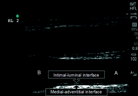
Fig. 10.2
Ultrasound image of the distal common carotid artery (A) and the carotid bulb (B). Arrows demarcate the intimal-luminal interface and the medial-adventitial interface which form the ultrasonographic borders (“double-line sign”) of the carotid intima-media thickness (CIMT)
High frequency ultrasound probes of 10–12 MHz or greater are required to produce images of sufficient resolution to allow for accurate and reproducible quantification of IMT.
The distal common carotid artery is regarded to be most suitable for accurate and reproducible measurement of carotid intima-media thickness [7].
Other segments of the carotid artery to include the internal carotid artery and bulb segment have been also studied. These sites are more commonly affected by focal plaque, but are more influenced by measurement variability due to arterial angulation.
The double-line sign refers to a relatively echodense intimal-luminal interface which overlies a more echolucent medial-adventitial interface, which together comprise the ultrasonographic appearance of the carotid intima-media thickness (Fig. 10.2).
Digital images are recorded in a cine-loop manner and transferred to a workstation where commercially available edge-detection software is used for CIMT quantification (Fig. 10.3):
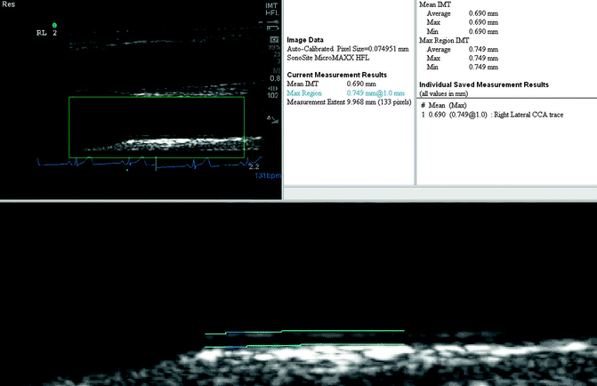
Fig. 10.3
An example of normal carotid intima-medial thickness measured utilizing edge-detection software (bottom inset). A mean CIMT of 0.690 mm is reported in this 66 year-old white male representing a value below the 50th percentile of CIMT distribution among healthy subjects classified according to age, gender and race
State of the art imaging requires that the arterial image be captured from a diastolic frame. This is to prevent phasic systolic wall thinning (of approximately 5 %) from influencing the measurements.
Measurement of the far-wall of the common carotid artery (furthest from the ultrasound probe) is preferred over near-wall measurement, which has been shown to be subject to more variation in gain settings, signal loss from acoustic shadowing from the relatively echo-bright adventitia, and limitations involving axial resolution. Far-wall CIMT determination by B-mode ultrasound has been shown to more accurately correlate with true arterial wall thickness in several studies [8–10].
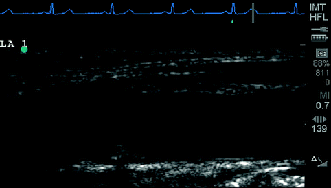
Sonographer experience and technical aspects can significantly affect reproducibility and accurateness of data collection. Figures 10.4, 10.5 and 10.6 illustrate some commonly encountered pitfalls and technical obstacles in image acquisition.
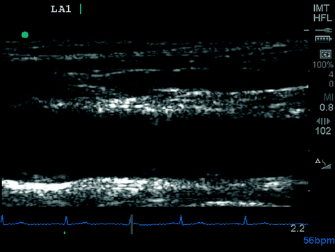
Fig. 10.4
Example of “near-wall artifact” which obscures the far wall luminal-intimal and medial-adventitial interfaces, preventing measurement of IMT. Optimizing gain can reduce artifact and improve image quality
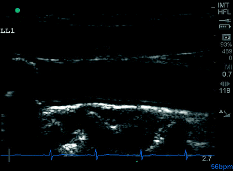
Fig. 10.5
Example of acoustic shadowing artifact. While visualization of intimal-luminal and medial-adventitial interfaces are preserved, this type of artifact can interfere with accurate measurement and adversely affect reproducibility
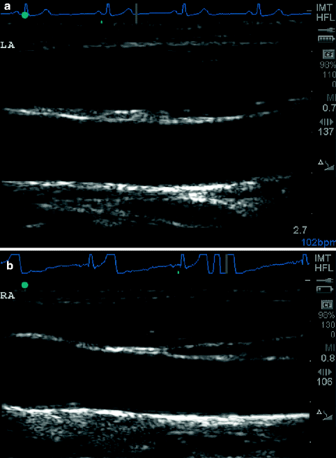
Fig. 10.6
(a, b) Examples of frequently encountered suboptimal carotid artery ultrasound imaging. Imaging plane too superficial or too deep resulting in absence of carotid bulb from image (a), and transducer not horizontally aligned with artery resulting in off-axis image (b). Both of these improper imaging techniques can result in inaccurate CIMT measurement
Methods to quantitate the degree of ultrasound “brightness” are available either through gray-scale quantification or through the quantification of the ultrasonic back-scatter characteristics. The use of back-scatter is less dependent on overall image gain and insonation angle but is more technically demanding. Histologic data suggest that echo-bright carotid atherosclerosis contains increased fibrous elements, whereas echolucent carotid atherosclerosis has increased lipid content.
3-dimensional, tomographic techniques are under development for volumetric quantification of carotid atherosclerosis.
Normal and Abnormal CIMT
CIMT values range within a healthy population according to age, gender, and race, and normal values for intima-media thickness have been defined based on this distribution [1, 11].
Multiple large population studies have demonstrated that intima-media thickness:
Normal CIMT is often considered to be the ≤75th percentile of CIMT distribution, a value which generally corresponds to an absolute value of CIMT of <1.0 mm (Fig. 10.3).
Focal plaque has been defined as a focal increase in CIMT > 1.5 times that of surrounding CIMT, or alternatively as areas of CIMT > 1.2 mm (Fig. 10.7).
Related posts:
 Insights into the Natural History of Atherosclerosis Progression
 Positron Emission Tomography: Imaging Inflammatory Atherosclerosis
 Factors Contributing to the Development of Atherosclerosis
 Intravascular Ultrasound
 Noncontrast Cardiac CT for the Detection of Calcified Atherosclerosis
 Coronary Atherosclerosis Imaging by Coronary CT Angiography

Stay updated, free articles. Join our Telegram channel

Full access? Get Clinical Tree


