Ascites
William J. Cochran
Ascites is derived from the Greek word “askos,” which means bladder or bag. Ascites is the accumulation of fluid in the peritoneal cavity; it is a manifestation of an underlying disorder, such as cirrhosis, congestive heart failure, nephrotic syndrome, protein-losing enteropathy, or malnutrition associated with hypoalbuminemia. Hippocrates stated, “When the liver is full of fluid and this overflows into the peritoneal cavity so that the belly becomes full of water, death follows.” Indeed, this was the case for Beethoven, who, in 1827, died 2 days after having a paracentesis performed. Although the predicted outcome is less bleak today, ascites resulting from cirrhosis still is associated with significant morbidity and mortality. In adults having cirrhosis and ascites, the 1-year survival is about 50%, compared with over 90% for those having cirrhosis without ascites.
PATHOGENESIS
The presumed initiating factor in the development of ascites in cases of congestive heart failure is increased hydrostatic pressure. In patients with the nephrotic syndrome, protein-losing enteropathy, or malnutrition, the associated hypoalbuminemia results in a decreased oncotic pressure. These alterations in Starling forces cause fluid to move from the intravascular space to the extravascular space. When the rate of extravascular fluid production exceeds the ability of the lymphatic system to reabsorb this fluid and transport it back to the vascular system, fluid accumulates in the peritoneal cavity, resulting in ascites.
The exact role of hypoalbuminemia in the development and maintenance of ascites is controversial, because many patients with hypoalbuminemia or analbuminemia do not have ascites. Approximately 50% of patients with serum albumin concentrations of less than 2.5 g/dL develop ascites. The pathogenesis of ascites in cirrhosis is less well defined and remains an area of active research.
In the past, two major theories had been proposed to explain the formation of ascites associated with cirrhosis: the underfill theory and the overflow theory. According to the underfill theory, ascitic fluid accumulates in the peritoneal cavity secondary to the alterations in Starling forces. Intrahepatic venous obstruction, caused by hepatic inflammation, scarring, and regenerative nodules, increases hydrostatic pressure in the hepatosplanchnic venous system. Increased hydrostatic pressure, in conjunction with a low oncotic pressure due to decreased hepatic protein synthesis, forces fluid out of the hepatosplanchnic vascular space and into the interstitial space. As the fluid in the interstitial space increases, eventually it exceeds the ability of the lymphatic system to reabsorb it, resulting in the accumulation of fluid in the peritoneal cavity as ascites. The intravascular volume is decreased, which stimulates the renin-angiotensin-aldosterone system to retain renal sodium to replenish the intravascular volume. Sodium retention increases the hydrostatic pressure in the hepatosplanchnic circulation, which promotes the accumulation of more ascitic fluid, establishing a vicious cycle.
Current evidence does not support this theory. If this theory were correct, vascular resistance should be increased along with a decrease in the cardiac index and plasma volume, which is not the case in patients with cirrhosis and ascites. More important, sodium retention has been well documented to precede the formation of ascites, rather than being the consequence of ascites.
The overflow theory proposes that the primary cause of ascitic fluid accumulation is renal sodium retention and subsequent plasma volume expansion. The sodium retention causes an expansion of the intravascular space, which increases the hydrostatic pressure in the hepatosplanchnic circulation and results in fluid extravasation into the peritoneal cavity. Several theories have been proposed to account for this increased sodium reabsorption, including decreased hepatic clearance of sodium-retaining substances or a reduced synthesis of a natriuretic substance. The major problem with this theory is that, instead of having an arterial vascular space that is overfilled, patients with cirrhosis and ascites actually have an arterial vascular system that is underfilled, even though the overall plasma volume and cardiac index are increased.
The most commonly accepted theory at this time is the arterial vasodilatation theory. This theory proposes that the initiating event is the development of peripheral arterial vasodilation resulting from an overproduction of vasodilators, primarily nitric oxide. The source of increased nitric oxide production is the endothelial cells of the splanchnic bed. In addition to overproduction, theoretically, diminished hepatic function results in the decreased inactivation of endogenous vasodilators, such as glucagon, vasoactive intestinal polypeptide, or substance P. The accumulation of endogenous vasodilators decreases the systemic vascular resistance, which decreases effective blood volume. This decrease in effective arterial blood volume is detected by arterial receptors, which in turn results in the stimulation of the renin-angiotensin-aldosterone system and the sympathetic nervous system, prompting renal sodium and water retention. The activation of the renin-angiotensin-aldosterone system increases renal vascular resistance and promotes the proximal and distal tubular reabsorption of sodium. In addition to increasing renal vascular resistance, activation of the sympathetic nervous system also promotes the proximal tubular reabsorption of sodium directly and increases renin secretion. This outcome increases the total blood volume, increasing the hepatosplanchnic circulation and its hydrostatic pressure, and resulting in fluid extravasation. Splanchnic arterial vasodilation occurs relatively early in the course of chronic liver disease. This finding is manifested clinically by a resting tachycardia and a wide pulse pressure found in persons with cirrhosis without ascites.
Several lines of evidence support this proposed mechanism of ascites formation. First, increased levels of multiple vasodilators have been noted in patients with cirrhosis and ascites. In addition, it has been determined that an increased production of nitric oxide occurs in patients with cirrhosis and ascites, as compared with normal individuals. Also supportive of this theory is the fact that the activity of the renin-angiotensin-aldosterone system and the sympathetic nervous system is increased in patients with cirrhosis and ascites. These patients have elevated levels of renin, antidiuretic hormone, and aldosterone.
Although the arterial vasodilation theory does not account for all the complex cardiovascular and renal changes in patients with cirrhosis and ascites, it is the explanation accepted most commonly. Additional research is needed to elucidate the pathogenesis of ascites in these patients.
DIAGNOSIS
The causes of ascites are subdivided into eight major categories: portal hypertension, hypoalbuminemia, infectious ascites, chylous ascites, urinary ascites, gastrointestinal ascites, miscellaneous causes, and pseudoascites (Box 365.1). Portal hypertension, the most common cause of ascites in North Americans, can have a prehepatic, hepatic, or posthepatic origin. The major cause of prehepatic portal hypertension is portal vein thrombosis or occlusion, which can result in the development of esophageal varices but rarely causes ascites. Often, hepatic-origin portal hypertension is secondary to hepatic fibrosis or cirrhosis. These disorders can result from congenital hepatic fibrosis, neonatal hepatitis, biliary atresia, alpha-1-antitrypsin deficiency, cystic fibrosis, chronic active hepatitis, or one of several storage diseases (see Chapter 370, Cirrhosis). Primary and metastatic hepatic tumors rarely may cause portal hypertension and ascites. Hepatic cysts, which may result in ascites, occur with polycystic kidney disease. Posthepatic causes of portal hypertension include the Budd-Chiari syndrome (i.e., hepatic vein thrombosis), constrictive pericarditis, or congestive heart failure. These latter two emphasize the importance of a thorough cardiac examination in the evaluation of a patient with ascites.
Hypoalbuminemia may be associated with ascites. The disorder associated most commonly with hypoalbuminemia and ascites is the nephrotic syndrome, although protein-losing enteropathy, malnutrition, and hydrops fetalis also can be responsible.
Ascites caused by infectious agents requires prompt diagnosis and treatment. Primary infections that may be associated with ascites include bacterial, fungal, or tuberculosis infections. Congenital cytomegalovirus, toxoplasmosis, or syphilis infections may be associated with significant ascites. Spontaneous bacterial peritonitis occurs in patients who have preexisting ascites and subsequent peritoneal infection by the hematogenous route. Patients with ascites may develop secondary bacterial peritonitis when a bowel perforation leads to peritoneal infection.
BOX 365.1 Categories in the Differential Diagnosis of Ascites
Portal hypertension
Prehepatic
Portal vein thrombosis or occlusion
Hepatic
Fibrosis
Cirrhosis
Tumors
Cysts
Posthepatic
Budd-Chiari syndrome
Constrictive pericarditis
Congestive heart failure
Hypoalbuminemia
Nephrotic syndrome
Protein-losing enteropathy
Malnutrition
Hydrops fetalis
Infectious causes
Bacterial peritonitis
Fungal peritonitis
Tuberculous peritonitis
Cytomegalovirus
Toxoplasmosis
Syphilis
Chylous causes
Traumatic
Lymphatic obstruction
Lymphatic abnormalities
Urinary causes
Posterior urethral valves
Bladder perforation
Ureteral stenosis
Urethral stenosis
Neurogenic bladder
Gastrointestinal causes
Pancreatic causes
Intestinal atresia
Meconium peritonitis
Bile peritonitis
Miscellaneous causes
Gynecologic disorders
Ventriculoperitoneal shunts
Eosinophilic peritonitis
Hypothyroidism
Pseudoascites
Omental cysts
Mesenteric cysts
Enteric duplication
Chylous ascites can be associated with trauma, lymphatic obstruction, or lymphatic abnormalities. Traumatic chylous ascites can result from a surgical procedure, an accidental blunt or penetrating injury, or child abuse. The most common cause of lymphatic obstruction that produces chylous ascites is lymphadenopathy. Neoplasms are a rare cause of chylous ascites in children, although they are the cause of chylous ascites found most commonly in adults. The major lymphatic abnormalities associated with chylous ascites are lymphangiectasia, lymphangiomatosis, and congenital “leaky lymphatics.” The latter disorder occurs in infants who are younger than 2 months and have chylous ascites of unknown cause. The disorder is thought to result from delayed maturation of the lacteals, allowing chyle to leak into the peritoneal cavity.
Urinary ascites results from leakage of urine into the peritoneal cavity. This process is responsible for approximately 50% of the cases of neonatal ascites. Posterior urethral valves cause approximately 60% of cases, and spontaneous congenital bladder perforation is responsible for 20%. Less common causes of urinary ascites are ureteral stenosis, urethral stenosis, and neurogenic bladder. Renal scintigraphy can be useful for localizing the area of leakage in these disorders.
Gastrointestinal disorders are an uncommon cause of ascites. Pancreatic ascites can be associated with pancreatitis or pancreatic pseudocysts. Pancreatic ascites is due to the contiguous spread of the pancreatic inflammation to the peritoneum, with extravasation of pancreatic secretions into the peritoneal cavity resulting in peritonitis. Rarely, neonatal ascites has a pancreatic origin. Other potential gastrointestinal causes of neonatal ascites are intestinal atresia, meconium peritonitis, and bile peritonitis.
Ascites rarely results from gynecologic disorders, such as ovarian cysts or pelvic inflammatory disease. Infrequently, ventriculoperitoneal shunts are associated with ascites. Eosinophilic peritonitis is a rare cause of ascites in children but is diagnosed readily from a markedly elevated eosinophil count in the ascitic fluid. Hypothyroidism may be associated with ascites, which resolves with thyroid replacement therapy.
Disorders that can mimic ascites, or pseudoascites, include omental cysts, mesenteric cysts, and enteric duplication. Failure to differentiate pseudoascites from true ascites before performing a paracentesis may be detrimental to the patient.
Physical Examination
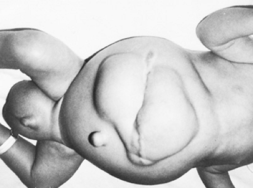
The clinical hallmark of ascites is abdominal distention (Fig. 365.1). Other potential physical findings include bulging flanks, protrusion of the umbilicus, and labial and scrotal swelling. Patients with portal hypertension may have a prominent abdominal venous pattern (Fig. 365.2).
Massive ascites (see Fig. 365.1) renders affected patients’ condition obvious. In less dramatic presentations, three physical signs can help to detect ascites: flank dullness, shifting dullness, and fluid wave. Flank dullness is verified with the patient in the supine position. In patients with ascites, the gas-filled loops of bowel float to the center of the abdomen, on top of the ascitic fluid. When the physician percusses the abdomen, it is tympanitic at the umbilicus and dull below the level of fluid into the flanks. Shifting dullness can be assessed by percussing the abdomen with the patient in the supine position and then in the right and left lateral decubitus positions. Ascites is suggested if the point of dullness shifts with changes in position. A fluid wave is elicited by having affected patients place the lateral aspect of their hands longitudinally on the abdomen. The examiner taps the lateral abdominal wall lightly while feeling the opposite wall for a fluid wave. Flank dullness and shifting dullness have the greatest sensitivity, and the fluid wave has the greatest specificity.
Previously, the puddle sign was advocated to test for ascites. After having been prone for 5 minutes, affected patients
rise on their hands and knees while the examiner lightly taps the flanks, auscultating the most dependent portion of the abdomen. The test is positive for ascites if a sloshing sound or change in the percussive sound occurs with lateral movement of the stethoscope.
rise on their hands and knees while the examiner lightly taps the flanks, auscultating the most dependent portion of the abdomen. The test is positive for ascites if a sloshing sound or change in the percussive sound occurs with lateral movement of the stethoscope.
 FIGURE 365.1. A 6-month-old infant with severe neonatal hepatitis. Note the marked abdominal distention, bulging of the flanks, wound and umbilical herniation, and scrotal swelling. |
The physical examination alone is not sufficiently specific or sensitive to detect ascites. In one study of patients with equivocal ascites, the physical examination alone had an accuracy of approximately 50%.
Patients in whom ascites is secondary to cirrhosis may have physical signs of chronic liver disease, such as large hemorrhoids, peripheral edema, scleral icterus, dilated abdominal vessels, spider telangiectasia, and splenomegaly. Other aspects of a physical examination that require particular attention in ascites patients are the cardiac and chest examinations. As discussed, several potential cardiac causes of ascites have been identified, including constrictive pericarditis and right ventricular failure. A thorough cardiac examination is required in all patients with ascites. The examination should include a search for jugular venous distention that suggests right ventricular failure. Some patients with right ventricular failure notice a pulsatile sensation on palpation of the liver. Patients with massive ascites may be tachypneic, owing to compromised intrathoracic volume. Some patients may develop sympathetic pleural effusions. The thyroid gland also should be palpated to evaluate for enlargement, which would suggest hypothyroidism.
Stay updated, free articles. Join our Telegram channel

Full access? Get Clinical Tree


