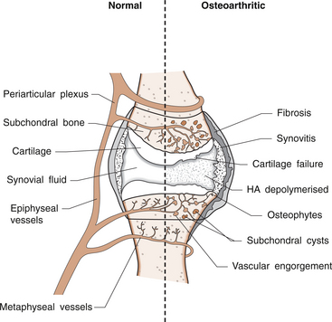
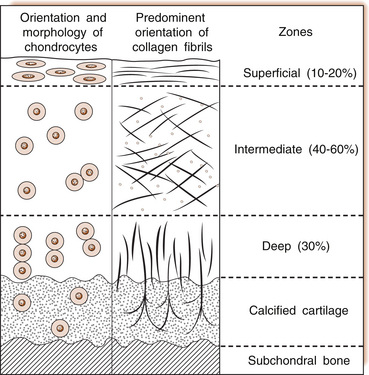
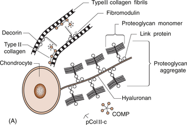
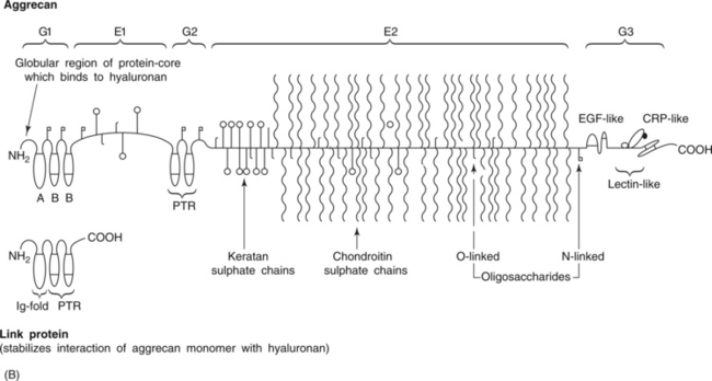
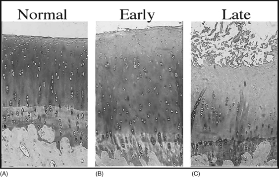
ARTICULAR CARTILAGE IN HEALTH AND DISEASE

6


ARTICULAR CARTILAGE IN HEALTH AND DISEASE


Lyn March and Chris Little


Introduction


The principal function of articular cartilage is to act as a protective shock absorber for the underlying bones. When it degenerates or becomes ‘diseased’, osteoarthritis ensues, with its accompanying pain and disability. Osteoarthritis affects approximately 10% of the population of the western world and is the leading cause of chronic disability in these communities. It increases dramatically with age, with as many as 50% of those over 65 years suffering from musculoskeletal symptoms, the majority of which will be due to osteoarthritis. No ‘cure’ as such exists for osteoarthritis but much is now known about risk factors that could contribute to prevention of progression, and numerous interventions, including non-pharmacological ones, have proven benefits for symptom relief.


This chapter will review normal structure and function of articular cartilage, as well as the normal anatomy, structure and function of the synovial joint with a focus on the knee joint. The ‘diseased’ state of articular cartilage describes the pathological process of osteoarthritis and a case will demonstrate the clinical symptoms and signs, risk factors and treatment of this condition.


Anatomy


The knee is a synovial joint with a surrounding capsule, an internal synovial lining that produces the lubricant joint fluid, internal menisci and stabilizing ligaments, and articular cartilage covering the surface of the bones. In the pathogenesis of osteoarthritis, changes occur in all structures including the synovial lining layer, the synovial fluid, the articular cartilage and the subchondral bone (Fig. 6.1). It is felt that the earliest and most significant changes occur in the articular cartilage and this chapter will focus on these.


Articular cartilage


Articular cartilage is a specialized form of connective tissue that covers and protects the ends of the bones in synovial joints. For the knee joint this makes up the smooth surfaces covering the femoral and tibial condyles and the under surface of the patella. The surface is smooth and slippery with an extraordinarily low coefficient of friction, while the deeper layer merges with a calcified layer (the tidemark) that interlocks with the subchondral bone (Fig. 6.2).


Cartilage is an elastic, resilient structure that acts as a shock absorber to protect the underlying bone. The properties of articular cartilage depend on the composition and structure of the extracellular matrix, and the synthesis and maintenance of this matrix is dependent on the chondrocytes.


During skeletal development, the articular cartilage forms from very densely packed mesenchymal cells that differentiate into chondrocytes, which proliferate rapidly and synthesize the large amounts of extracellular matrix. The extracellular matrix is made up predominantly of water (up to 80%), collagen and proteoglycans (discussed below), which are produced and maintained by the relatively sparse cells, the chondrocytes. It is the combination of collagen, proteoglycan and water that gives articular cartilage its unique properties. The collagen forms a network of fibrils that gives the overall framework and shape of the cartilage and provides pockets or compartments that are filled with the water-binding proteoglycan complexes that regulate the compressibility. The ability of articular cartilage to resist compressive deformation is largely due to the entrapment of high concentrations of the polyanionic large proteoglycan aggrecan within the collagen fibrillar network. The osmotic pressure provided by the glycosaminoglycan (GAG) chains on the aggregated aggrecan molecules is resisted by and constrained within the insoluble collagenous meshwork. Degradation of cartilage is a central pathological feature of arthropathies such as osteoarthritis and rheumatoid arthritis, and involves proteolytic cleavage of both its major structural elements, namely aggrecan and type II collagen. Proteolysis and subsequent loss of the GAG-rich region of aggrecan from cartilage is an early event in cartilage degeneration, while significant catabolism of the collagen fibrillar structure occurs later and may represent the point of irreversible cartilage damage.


The functional integrity of the articular cartilage in a healthy joint depends on the chondrocyte synthesizing the many different matrix components in the appropriate amounts and in the right sequence. Since cartilage lacks blood and lymphatic vessels, the survival and synthetic activity of the chondrocyte depends on the diffusion and transport of nutrients and metabolites through the matrix. Thus a fine balance exists between anabolism (synthesis) and catabolism (tissue breakdown) with ongoing tissue remodelling part of the normal healthy process.


In ‘disease’, osteoarthritis, this equilibrium is disturbed with the balance tipped in favour of degenerative changes and the chondrocyte failing to keep up, despite an observed initial increase in chondrocyte numbers and synthetic processes.


The chondrocyte


Chondrocytes play a unique role in regulating both synthetic and catabolic processes in health and disease. They make up less than 5% of the total volume of the cartilage. Each chondrocyte establishes a specialized microenvironment and is responsible for the turnover of extracellular matrix in its immediate vicinity. Cells in different parts of the tissue appear to have metabolic differences, because different components of the matrix are required to maintain structure and function.


Chondrocytes vary in shape, size and number of cells per area depending on their different anatomical locations, even within different regions of the normal knee joint. Close to the surface they are flatter, smaller and generally have a greater density than the cells deeper in the matrix. Each chondrocyte sits within a lacuna (space). Collagen fibres come right up to the edge of the lacunae, which are filled with fine fibrillar material. Unlike the osteocyte in bone, their cytoplasmic processes do not make contact with processes of other chondrocytes i.e. they have little or no cell-cell contact. They have low numbers of mitochondria, which reflects their low oxygen consumption rates. In adult cartilage, the rate of cell division is very low but division does occur in response to injury or disease. Chondrocytes, particularly in the deeper uncalcified zone, have prominent endoplasmic reticulum and Golgi apparatus responsible for protein synthesis and sulphation of the mucopolysaccharides that form the proteoglycan side-chains. Lipid occurs intracellularly and as a diffuse layer around the cells, and probably contributes to cartilage lubrication.


A number of in vivo and in vitro studies have shown that environmental changes can alter chondrocyte function and thus the surrounding matrix. There appears to be an optimal window of mechanical loading. At low levels of mechanical stress, as would occur with bed rest or disuse, there is increased catabolic activity; at physiological levels there is anabolic activity; while at higher stress levels, as would occur with obesity or carrying heavy loads repeatedly, the chondrocyte is unable to adapt and catabolic processes outstrip the anabolic activity.


Biochemistry


Collagen network


There are at least 13 different types of collagen in the connective tissues throughout the human body. In articular hyaline cartilage, the predominant components are types II, IX and XI with small amounts of others.


Type II collagen is the predominant fibrous component making up 90–95% of the primary collagen and 40–70% of the total dry weight of articular cartilage. It forms a three-dimensional cross-banded network of fibrils that maintain the ‘shape’ of the cartilage structure. It is secreted by the chondrocytes as procollagen molecules that consist of polypeptide chains organized as a triple helix. These grow and aggregate to form fibrils that then develop strong covalent interfibrillar cross-links. The minor collagens, such as types IX and XI are thought to be important for regulating structure and interactions.


In osteoarthritis, collagen network swelling has been noted as one of the earliest features of the fibrillation process. The chondrocyte is known, when stimulated by inflammatory mediators such as the cytokine interleukin-1 (IL-1), to produce enzymes called metalloproteinases (including stromelysin and collagenases) that degrade the collagen molecules into fragments. This contributes to the loosening of the collagen network and allows the increased water content that is also a feature of early osteoarthritis.


Proteoglycans


Proteoglycans are the predominant molecules trapped in the collagen fibrils and make up 15–40% of the dry weight of articular cartilage. They have many negative charges and thus are highly hydrophilic, which leads to trapping of water which, in turn, contributes to the shock-absorbing capacity.


Proteoglycans are made up of a core protein with glycosaminoglycan (GAG) side-chains; GAGs are linear polysaccharides made up of many repeating disaccharide units—including keratan sulphate (KS) and chondroitin sulphate (CS)—together with numerous other oligosaccharides.


Most proteoglycans exist in the cartilage matrix as aggregates. As many as 100 proteoglycan monomers will lock onto a central hyaluronic acid (HA) filament, stabilized by link protein (Fig. 6.3). Their aggregate molecular weights are enormous—1 to 2 million daltons (Da).


Proteoglycans play a crucial role in the ability to absorb loading forces in a reversible way. They are responsible for restricting water flow and thus resist deformation during compressive loading. As the tissue is compressed, e.g. in weight bearing, some water is squeezed out, but much is held in by the attractive forces of the proteoglycans; the proteoglycans come into closer contact but are repelled by their strong charges, preventing deformation. As the load is removed, the tissue rapidly regains its form by taking on the water again.


The highly charged state of proteoglycans also stops the flow of large molecules across the tissue but allows the smaller molecules to diffuse through, which is an important mechanism for delivering nutrients to the avascular cartilage.


Pathophysiology of osteoarthritis


When the delicate balance of cartilage matrix synthesis and destruction is upset, the net result is articular cartilage loss and the degenerative process that ensues is osteoarthritis. Despite significant advances in our understanding of cartilage structure, there is still a lot to learn about the mechanisms of cartilage destruction. While it is felt that the initial pathology in osteoarthritis involves the articular cartilage, it is evident from very early stages that there are changes also in the synovial lining, the synovial fluid and the underlying subchondral bone.


Articular cartilage


Macroscopically, the cartilage surface undergoes three phases of degeneration starting with fibrillation and progressing through to erosion and cracking to expose the underlying subchondral bone. Eventually, the bone surface may be denuded of its cartilage cover, a process termed eburnation. In the knee joint, the areas of cartilage damage are most apparent on the surfaces exposed to excessive load bearing and may be quite focal in the early phases.


At the biochemical level, the main changes in osteo-arthritis involve an increase in the water content of the articular cartilage, a decrease in the proteoglycan concentration and loss of the collagen network. In the early phases, the articular cartilage will actually thicken and swell because of the increased water and early increased synthesis of proteoglycans. However, these changes leave the cartilage less compressible, more permeable to tissue breakdown products and thus more prone to damage from impact loading. The collagen network also breaks down as enzymes are released from stressed chondrocytes and synovial lining cells. The enzymes belong to a family called metalloproteinases, of which collagenase is a member. The cartilage softens (termed chondromalacia) and progresses to fibrillation of the surface layers.


Microscopically, there is evidence of chondrocyte necrosis as well as focal clumps or clones of increased proliferation. The proliferating cells are attempting repair and have been shown to increase proteoglycan production locally. The ‘repaired’ cartilage tends to be more cellular and, at points where erosions have occurred and bone has been denuded, it has the properties of fibrocartilage rather than the resilient hyaline cartilage of a normal healthy joint. The main collagen type in fibrocartilage is type I rather than type II and the increased fibrous content offers less protection to the underlying bone.


Pathology occurs in all joint tissues in OA, including bones, menisci, ligaments and synovium but the central feature is progressive degeneration of the articular cartilage (Fig. 6.4). Articular cartilage has a very poor reparative capacity, and ultimately it is the breakdown and erosion of this tissue that signifies end-stage arthritis necessitating joint replacement surgery.


What mechanisms are important in cartilage breakdown?


Cartilage is aneural, avascular, sparsely populated with cells (chondrocytes) and predominantly comprised of extracellular matrix (Fig. 6.4A). The major matrix proteins are type II collagen which forms a structural scaffold, and aggrecan (the aggregated proteoglycans) which is substituted with negatively charged glycosaminoglycans. The swelling pressure of the aggrecan is balanced by the tension in the collagen network, and together they endow cartilage with its resilience under compression. Progressive breakdown of cartilage in OA involves proteolysis of both aggrecan and type II collagen. Aggrecan breakdown precedes and is independent of collagen degradation, and both in vitro and in vivo is due to the action of members of the A Disintegrin And Metalloproteinase with ThromboSpondin motif (ADAMTS) family of enzymes. In particular ADAMTS-4 and -5 appear to be the physiologically important ‘aggrecanases’. Although cartilage has a limited ability to repair, aggrecan can be replenished by the chondrocytes and the mechanical properties of the tissue restored if the destructive insult is removed prior to disruption of the collagen network. Once significant disruption of the collagen network occurs (Fig. 6.4C), cartilage damage is irreparable with further collagen breakdown resulting in progressive cartilage loss.


Fibrillar type II collagen is resistant to degradation by all proteinases at 37°C and neutral pH, except the collagenolytic metalloproteinases (MMPs) (MMP-1, -2, -8, -13 and -14). It has been shown that cytokine-stimulated collagenolysis in articular cartilage is due to the action of MMPs with MMP-13 being the principal enzyme both in vitro and in vivo.


Activation of MMPs in cartilage


MMPs are secreted as zymogens and must be activated extracellularly where their activity is then controlled by tissue inhibitors of metalloproteinases (TIMPs). Chondrocyte mRNA levels for numerous MMPs including MMP-2, -9 and -13 are significantly increased in OA cartilage in both animals and humans. While OA is often considered a ‘non-inflammatory’ arthropathy, chondrocyte-derived cytokines such as IL-1 and TNF are known to play a direct role in human cartilage degradation by MMPs. However, the precise regulatory pathway whereby chondrocyte MMP expression is regulated, and therefore the identification of potential therapeutic targets, is still the subject of intensive investigation.


Subchondral bone


As the articular cartilage is eroded, the underlying bone becomes exposed to wear and tear, causing a polishing effect on the surface and microfractures of the bony trabeculae. In response, there is increased osteoblastic activity and new bone formation. The surface may also undergo focal pressure necrosis. As the overlying cartilage is denuded, subarticular cysts may develop. This is thought to be due to the combination of the focal necrosis and the transmission of intra-articular pressures through to the marrow spaces of the underlying bone. Cysts can collapse or may regress or even disappear if the surface becomes covered with regenerative fibrocartilage. Vascular engorgement, slowing of blood flow and bone marrow oedema in the subchondral bone are all features documented at different stages of the osteoarthritis cycle and are thought to contribute to some of the clinical features of pain.


Synovium


As discussed in Chapter 1, the normal synovium consists of two layers: the lining layer of cells, known as the intima, and the remaining subsynovial tissue or subintima. The normal intima is formed by an interlacing layer, one to three cells (synoviocytes) deep, that merges with the underlying connective tissue. There is no basement membrane between. The lining is generally smooth with few folds and the subintima has connective tissue cells (fibroblasts) only and relatively few blood vessels and endothelial cells.


In osteoarthritis, products released from the breakdown of the cartilage and bone evoke an inflammatory response in the synovium, which becomes both hyperplastic, with increased numbers of synovial lining cells, and hypertrophic, with small villi or folds developing in the membrane, infiltration of lymphocytes and plasma cells and an increase in the vascularity in the subintimal layer. This is in contrast to rheumatoid arthritis where the synovial inflammation is thought to be the primary pathology, leading to secondary cartilage destruction in which the synovial proliferation is much more marked. One of the functions of the synovial membrane is to provide nutrients for the avascular articular cartilage, but if thickened and scarred, it may not function as well and thus may perpetuate the degenerative cycle. The influx of inflammatory cells releases mediators that further contribute to tissue damage.


Synovial fluid


Normal synovial fluid is clear, pale yellow in colour and very sticky or viscous (see Ch. 1). It is a dialysate of plasma combined with hyaluronic acid that is produced by the synovial cells. The molecular weight of HA determines the elasticity and viscosity of the fluid. A normal knee joint contains 0.5–1.5 mL of fluid, which coats the surface of the articular cartilage, providing lubrication for movement, a vehicle for flow of nutrients from the fluid into the articular cartilage matrix and a shock-absorbing cushion on weight bearing. In OA there is often an increased volume of fluid but the hyaluronic acid content and thus the viscosity and other mechanical properties of the fluid are reduced. The fluid is still usually clear and non-inflammatory in OA but if cartilage fragments and/or low-grade inflammation are present, it will have a more turbid appearance.


Normal fluid may produce a ‘string’ sign, in which the fluid drop can hang from a thread of synovial fluid owing to the high viscosity. The greater the level of inflammation, the more fluid and the less viscous it becomes. Normal synovial fluid has very few white cells (<100/mm3) and fluid from osteoarthritis is also characteristically non-inflammatory. Cell counts may be slightly higher (100–2000/mm3) but cells will be predominantly mononuclear, unlike in rheumatoid arthritis where cell counts are much higher (>5000/mm3) with a higher percentage of polymorphonuclear cells. Higher levels of cells may be associated with inflammation due to calcium pyrophosphate or hydroxyapatite crystals that may be present at sites of tissue damage. Increased white cell numbers may, in turn, be associated with accelerated tissue damage because of the release of inflammatory mediators.


Clinical features of osteoarthritis


Symptoms and signs of osteoarthritis are summarized in Box 6.1.


Jul 3, 2016 | Posted by in MUSCULOSKELETAL MEDICINE | Comments Off on ARTICULAR CARTILAGE IN HEALTH AND DISEASE

Full access? Get Clinical Tree

Get Clinical Tree app for offline access