(1)
Department of Orthopedic Surgery Academic Medical Centre, University of Amsterdam, Amsterdam, The Netherlands
Abstract
This chapter describes the indications and operative technique of arthroscopy of the first metatarsophalangeal joint. The main indications are impingement, hallux rigidus, and osteochondritis dissecans. Using the four portals described, it is possible to visualize the entire first MTP joint.
The main indications are impingement, hallux rigidus, and osteochondritis dissecans.
26.1 Introduction
Watanabe et al. described the first arthroscopy of the first metatarsophalangeal (MTP) joint in 1985 (Watanabe et al. 1985): “The articulation between hallux and its metatarsal is entered at the point either lateral or medial to the extensor tendon. As this joint is extremely difficult to open with manual traction, a 2.7 mm outside diameter sheath must be used.” Bartlett reported a case in 1988 in which successful arthroscopic debridement was performed on an osteochondral defect (Bartlett 1988).
A series of 20 patients with arthrosis of the first MTP joint was reported in which diagnostic arthroscopy was performed. Shaving gave some promising results. However, it was not the osteoarthritis that made me decide to perform arthroscopy on the first MTP joint. Members of the Dutch National Ballet and other ballet groups often confronted me with pain problems and painful limitation of dorsiflexion in the first MTP joint. As with the anterior impingement syndrome of the ankle joint, the most likely cause is repetitive microtrauma on the dorsal aspect of the joint with secondary spur formation. Extension osteotomy of the first phalanx was the original approach to the problem. Because arthroscopic treatment of the anterior impingement syndrome of the ankle joint is highly successful, I tried the same procedure in the first MTP joint. The arthroscopic procedure has the advantage of less morbidity, outpatient treatment, and reduced risk of range of motion limitations that may be produced by the scarring that results from classic arthrotomy. Another advantage is faster return to sports and work (Van Sterkenburg et al. 2009).
These advantages hold for every patient; therefore, other indications were explored (Stroink and Van Dijk 2009).
26.2 Indications
The main indications for arthroscopy of the first metatarsophalangeal joint are dorsal impingement, osteochondritis dissecans, and hallux rigidus.
26.2.1 Impingement Syndrome
Dorsal impingement on the big toe is the result of repetitive microtrauma on the dorsal aspect of the joint. It results from repetitive forced dorsiflexion movement, as occurs with ballet dancing (Biedert 1991). This results in damage to the cartilage rim on the dorsal aspect of the distal phalanx and metatarsal head. An inflammatory reaction, synovitis, scar tissue formation, calcifications, and finally spur formation can be the result.
Clinically, there is pain on the dorsal and dorsolateral aspect of the joint, especially during or after forced dorsiflexion. In more severe situations, limitation of dorsiflexion evolves.
On examination, there is tenderness on the dorsal aspect of the first MTP joint, where a rim can be palpated. Typically, it is located at the dorsolateral aspect of the metatarsal head. Dorsiflexion is slightly limited. On X-ray there are no degenerative changes on the anteroposterior (AP) or lateral views. On the lateral view there is a spur beginning on the dorsal aspect at the junction of the metatarsal head and the shaft. The condition is comparable to the anterior impingement syndrome of the ankle joint. Here, we find the same painful limitation due to scar tissue and sometimes spur formation, usually without joint space narrowing (Van Dijk et al. 1995).
26.2.2 Osteochondritis Dissecans
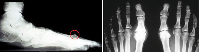
Osteochondritis dissecans in the first MTP joint is a well-known pathology (Carell and Childress 1940). Osteochondritis dissecans can lead towards a hallux rigidus deformity. The symptoms and therapy are no different from osteochondritis dissecans in other joints such as the ankle or knee joint. In some patients there is intermittent limitation of motion due to locking, caused by a loose body (Fig. 26.1).
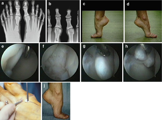
Fig. 26.1
A professional ballet dancer with intermittent locking of the first MTP joint. Since 3 months the first MTP joint is locked preventing him from full dorsiflexion. The picture shows the normal “demi point” position of the right foot (c) and the abnormal position of the left foot (d), with diminished dorsiflexion of the first MTP joint. The X-ray (a, b) shows no abnormality except for a possible shallow defect (arrow) in the medial tarsal head (left ankle). Arthroscopy was performed (e–h) and a chondral loose body was found. After removal (i) the patient was symptom-free. We see the normal “demi point” 5 days postoperatively (j)
26.2.3 Sesamoiditis
The sesamoid bones are incorporated in the short flexors of the big toe and articulate with the plantar surface of the head of the metatarsal. A normal sesamoid bone can consist of multiple fragments (Kilokuim 1965).
Differentiation between a multifragmented sesamoid bone or a fracture/nonunion can be made by technetium bone scan. A fracture may be the result of trauma, but more often complaints in this region are the result of repetitive microtrauma. A fatigue fracture may be the ultimate result. The pain, located on the plantar side of the head of the first metatarsal, is present during or after activity. It can be elicited by forced dorsiflexion of the big toe. There is local pressure and pain but usually no swelling. AP and lateral views show the irregular, sometimes dislocated fragments. The axial view can demonstrate an irregular joint line between the sesamoid bone and the metatarsal head. A bone scan differentiates between active pathology and normal anatomy. Conservative treatment consists of a special inlay, anti-inflammatory drugs, plaster casting for a minimum of 6 weeks, and infiltration. Operative treatment with removal of the deformed sesamoid bone gives inconsistent results.
26.2.4 Hallux Rigidus
Keetley (1887) was the first to describe hallux rigidus (Fig. 26.2). Its possible etiologic factors are arthrosis deformans, repetitive microtrauma during sports activities (Waterman 1927), macrotrauma, aseptic necrosis, osteochondritis dissecans (Kingreen 1933; Goodfellow 1966; Hackenbroch 1927), and limited range of motion whereby dorsiflexion is progressively diminished. Pain finally makes normal walking impossible. An X-ray during the first stage may show no deformity. At a later stage, however, joint space narrowing and osteophytes become apparent. Operative options are resection of osteophytes, partial resection of the proximal one-third of the first phalanx (Brandes 1929), arthrodesis, and silicon interposition arthroplasty (Weirgart and Klems 1976; Hohlmann 1956; Van Dijk et al. 1998).