25 Arthroscopic “Wafer” Procedure
Indications
Pitfall
Cannot be used if positive ulnar variance is greater than 4 mm or when there is arthritis of the distal radioulnar joint (DRUJ)
Technique
- Standard wrist arthroscopy setup is used.
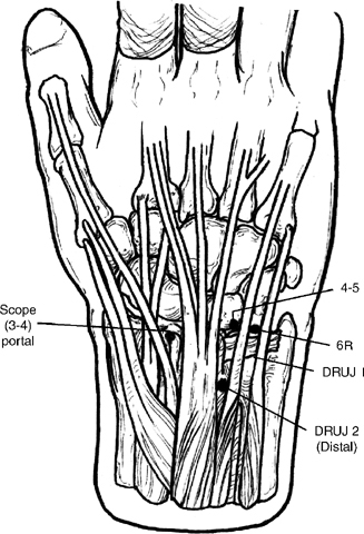
- Use the 3–4 and 4–5 or 6R portals to inspect the joint, specifically evaluating the triangular fibrocartilage complex (TFCC), lunotriquetral interosseous ligament, lunate, triquetrum, and ulnar head for tears and chondromalacia (Fig. 25-1).
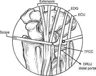
- Debride the central two thirds of the TFCC using a shaver, punch, or radiofrequency device in the 4–5 or 6R portal (Fig. 25-2).
Figure 25-1
Figure 25-2
Pitfall
Do not violate the peripheral 2 mm of the articular disk to avoid creating DRUJ instability.
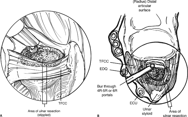
- Using a motorized bur in the 4–5 or 6R portal, remove the central portion of the ulnar head dome through the hole in the TFCC to create 2 mm ulnar negative variance (Fig. 25-3A,B).
- A distal DRUJ portal is made 3 mm ulnar to the sigmoid notch and just proximal to the TFCC.
Pearl
To make the DRUJ portal, position the forearm in slight pronation and spread a hemostat horizontally between the ulnar head and TFCC.
- Place the bur in the DRUJ portal and remove the periphery of the dome to complete the resection (Fig. 25-4).
- Pronate and supinate the forearm to reach the entire periphery of the dome.
Pitfall
Do not violate the fovea of the ulnar head where the deep fibers of the radioulnar ligaments attach.
- With wrist traction reduced, confirm a complete and adequate level of resection with fluoroscopy.
- Modify the resection level if necessary and remove any remaining sharp bony edges.


