Arthroscopic Treatment of the Degenerative Knee
J. Richard Steadman
The usefulness of arthroscopic treatment of osteoarthritis of the knee was recently challenged after one article showed there was no benefit when it was compared with irrigation or placebo.1 Although this article was statistically well constructed, it did fail to address several issues that, in my opinion, are crucial to the success of arthroscopic treatment of osteoarthritis of the knee. There are many characteristics in the degenerative knee that can produce pain. These include stiffness, synovitis, loose bodies, meniscus tears, and closed spaces due to compartmentalization, or old adhesions. Also, chondral defects or flaps can cause pain. In my experience, painful symptoms decrease about 70% of the time if these conditions are treated arthroscopically. Many of the studies have used arthroscopic techniques that include lavage, debridement, and abrasion arthroplasty.2,3,4,5,6,7,8,9,10,11,12,13,14 However, these studies have not focused on increasing joint volume, treating capsular contracture, and performing synovectomy. Furthermore, most of the studies have not emphasized the importance of the role of postoperative rehabilitation. Correct rehabilitation is essential to address stiffness, maintain joint volume, and achieve maximum range of motion. Strengthening exercises also protect the joint by improving concentric and eccentric muscle contraction. The goal of my arthroscopic treatment regimen is to allow patients to resume their active lifestyles, decrease their disability, and delay total knee arthroplasty.
Initial Evaluation
Patients may present with a variety of conditions. Many patients have malalignment, joint stiffness, a prior meniscectomy, and/or instability with stiffness. Symptoms include pain, swelling, stiffness, and mechanical symptoms. In the degenerative knee, pain generators include stiffness, synovitis, loose bodies, meniscus tears, and stiffness due to closed spaces from compartmentalization or old adhesions. Loss of patellar mobility is common in the knee with degenerative joint disease. This is usually accompanied by diminished range of motion, particularly in extension. Loss of articular cartilage in the degenerative knee may not cause pain if these other conditions are corrected.
Radiographic evaluation includes standing anteroposterior, 45 degree flexion posteroanterior, long standing, and patellar views. The long standing view from hip to ankle allows for the evaluation of malalignment, and the 45 degree flexion view will show any diminished joint
space posteriorly. Evaluating joint malalignment is discussed further in Chapter 13.
space posteriorly. Evaluating joint malalignment is discussed further in Chapter 13.
Patient Selection
This arthroscopic treatment protocol is appropriate for all age groups. It is usually considered after nonoperative measures such as weight loss, strengthening, medications, icing, and flexibility have not been successful in relieving pain. A knee sleeve can be helpful, but unloading braces are more effective if they are comfortable, well tolerated, and do not slip. Activity modification, such as changing from jogging to cycling or water exercise, is another option to consider. Additionally, injection with a cortisone preparation or viscosupplementation with a hyaluronic acid solution can be useful.
Treatment Package
My surgical treatment regimen for knees with severe osteoarthritis focuses on increasing joint volume with arthroscopy, and maintaining it with rehabilitation to provide symptomatic relief. This relief occurs when joint contact pressures are decreased, and correct rehabilitation keeps the joint spaces open. The comprehensive arthroscopic regimen consists of distinct procedures, which are performed after general anesthesia or spinal anesthetic is administered. A tourniquet is rarely used.
The first step in the procedure is to expand the joint space using insufflation. In the severely degenerative knee, the joint volume is contracted, with a volume of only 60 to 90 mL, whereas a normal knee has about 180 mL of volume. An 18-gauge needle and a 60-mL syringe are used to inflate the joint with a saline solution (Fig. 12.1).
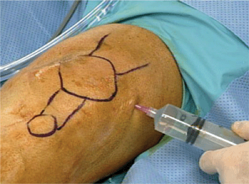 Figure 12.1 Needle placed in the suprapatellar pouch for the lateral side of the knee to inject fluid. A large needle is used to allow for tactile feedback to ensure that the fluid is flowing freely into the true joint space. |
TIP: It is important to make certain the needle is in the joint, and not in the subcutaneous tissue. Great care should be taken to avoid the back wall of the pouch. If the joint is entered superolateral to the patella and the needle is angled down, it is more likely to enter the joint and avoid injecting the back wall of the pouch. There should be an easy flow of solution into and out of the joint.
Fluid is inserted under manual pressure. As pressure increases, the outflow of fluid, when the syringe is removed from the needle, progresses from a drip to a steady flow, and then to a strong stream (Fig. 12.2). The joint is not like a balloon. If it is stretched, it will not just spring back. The degenerative joint has soft tissue contracture, and once it is stretched the resulting increased volume is maintained by fluid inflow during surgery, followed by mobilization and range of motion after surgery.
Related posts:
 The ACL-Deficient Knee: Some Thoughts—Past and Future
Imaging of the Knee
The Healing Response Technique: A Minimally Invasive Procedure to Stimulate Healing of Anterior Cruciate Ligament Injuries Using the Microfracture Technique
Functional Biomechanics of Healthy, Anterior Cruciate Ligament-Deficient and -Reconstructed Knees
Microfracture
The ACL-Deficient Knee: Some Thoughts—Past and Future
The ACL-Deficient Knee: Some Thoughts—Past and Future
Imaging of the Knee
The Healing Response Technique: A Minimally Invasive Procedure to Stimulate Healing of Anterior Cruciate Ligament Injuries Using the Microfracture Technique
Functional Biomechanics of Healthy, Anterior Cruciate Ligament-Deficient and -Reconstructed Knees
Microfracture
Stay updated, free articles. Join our Telegram channel

Full access? Get Clinical Tree


