The most critical step in successful treatment of shoulder instability does not lie in surgical technique, but in accurate assessment of factors contributing to instability. Multidirectional instability (MDI) is initially treated with rehabilitation. The primary goal of rehabilitation is strengthening of the dynamic stabilizers, including the rotator cuff and scapular stabilizers. There are several surgical techniques described to manage MDI, ranging from the classic Neer inferior capsular shift to a variety of arthroscopic procedures. This article focuses on the arthroscopic management of MDI.
In 1980, Charles S. Neer provided the first description of multidirectional instability (MDI) and the operation, the open inferior capsular shift, which was established as the gold standard. He described it as “involuntary inferior and multidirectional subluxation and dislocation.” This original article outlined the basis for much of our modern thinking about this condition. Although it is surprising how little has changed during the past 30 years, one aspect of MDI that has altered our thinking significantly is the advent of arthroscopic surgery and the innovation of arthroscopic techniques to restore stability and minimize morbidity.
Shoulder instability is best understood as a spectrum of disease. The most common type of instability, traumatic unidirectional instability, lies at one end of the spectrum, while multidirectional instability lies at the other extreme. Many cases fall somewhere in between these two classic examples. The most critical step in successful treatment of shoulder instability does not lie in surgical technique, but in accurate assessment of factors contributing to instability. Diagnostic precision is confounded further by variations in the definition of multidirectional instability. In general, MDI consists of symptomatic, involuntary instability of the glenohumeral joint in more than one direction. It is often bilateral and usually atraumatic. One of the frequent findings in MDI compared with traumatic unidirectional instability is pathologically increased capsular volume. MDI should not be confused with asymptomatic hyperlaxity or voluntary instability.
MDI is initially treated with rehabilitation, which requires patience and an extended period of time and effort on the part of the patient. The primary goal of rehabilitation is strengthening of the dynamic stabilizers, including the rotator cuff and scapular stabilizers. Proprioceptive training is also important. Nonoperative management is successful in approximately 80% of compliant patients with MDI. In the recalcitrant case in which nonoperative treatment fails, surgical management is appropriate. There are several surgical techniques described to manage MDI, ranging from the classic Neer inferior capsular shift to a multitude of arthroscopic procedures. This article focuses on the arthroscopic management of MDI.
Anatomic considerations
Shoulder stability is imparted by a combination of static and dynamic stabilizers. The most important static stabilizers include the glenohumeral ligaments, which are thickenings in the glenohumeral joint capsule that tighten and relax depending on the position of the shoulder. As a group, they render the shoulder stable through the full range of motion. The inferior glenohumeral ligaments (anterior and posterior) form a sling beneath the glenohumeral joint that stabilizes the joint in positions of abduction, preventing anterior, posterior, and inferior translation. The middle glenohumeral ligament provides anterior stability in the midrange of abduction, limiting external rotation and inferior translation. The superior glenohumeral ligament and coracohumeral ligament stabilize the shoulder in the adducted position, primarily limiting inferior translation and external rotation. These ligaments also comprise the structural portion of the rotator interval, the area of capsule between the superior edge of the subscapularis tendon, and the anterior edge of the supraspinatus tendon. Incompetence of the rotator interval has been shown to result in a 50% increase in posterior translation and a 100% increase in inferior translation.
The most important dynamic stabilizers include the rotator cuff muscles, scapular stabilizers, and the long head of the biceps. The rotator cuff is able to resist humeral head translation through the mechanism of concavity-compression, in which the humeral head is centered into the glenoid and the rotator cuff imparts a balanced contact pressure to the articulation. The labrum increases the concavity of the socket by up to 50%, but only provides 20% of the concavity-compression stability of the glenohumeral joint. Depth of the socket (including sufficient glenoid bone stock and labral integrity) and adequate compressive force (including intact rotator cuff and sufficient rotator cuff strength) are important for shoulder stability.
Scapular stabilizers are shown to be important for shoulder stability, and abnormal scapular kinematics and periscapular muscle function have been identified in patients with MDI. Decreased upward rotation and increased internal rotation in scapular plane abduction were found in patients with MDI in comparison with asymptomatic control patients. Stabilizing the scapula results in stabilization of the glenoid, the platform on which the humeral head must function.
Making the diagnosis
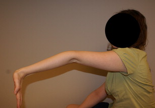
There are several potential causes of MDI. Genetic disorders such as Ehlers-Danlos syndrome can result in abnormal connective tissue properties that are present from birth and cannot be changed. Repetitive microtrauma such as can occur with swimming, throwing, or gymnastics can cause capsular damage that builds up over time. Patients with generalized ligamentous laxity are especially prone to develop MDI. Signs of laxity include elbow or knee hyperextension beyond 10°, small finger metacarpophalangeal hyperextension more than 90°, or the ability to abduct the thumb to the forearm with the wrist fully flexed. If the patient has 3 out of 4 of these signs, he or she is believed to have generalized ligamentous laxity ( Fig. 1 ). Skin hyperelasticity should also be noted. If severe generalized ligamentous laxity is found, a genetic workup for Ehlers-Danlos syndrome or other connective tissue disorders may be warranted. Patients who voluntarily dislocate their shoulders for reasons of psychiatric pathology or for secondary gain must be identified. These patients typically have poor results after surgery.
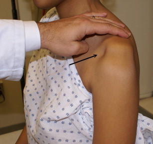
Patients may present with frank instability or with nebulous descriptions of pain exacerbated by positions that provoke instability such as throwing (anterior instability), carrying heavy loads (inferior instability), or pushing (posterior instability). Physical examination maneuvers for anterior instability include the apprehension, relocation, and anterior release tests, the load and shift test, and the anterior drawer test. Tests for posterior instability include the posterior stress test, the jerk test, the load and shift test, and the posterior drawer test. The most important maneuver in the MDI examination, however, is the sulcus test, initially described by Neer and Foster. Inferior traction is placed on the affected limb with the arm adducted at the side in neutral rotation. A positive finding is the presence of a dimple in the lateral subacromial aspect of the shoulder ( Fig. 2 ). This maneuver is repeated with the arm in 30° external rotation to determine whether the rotator interval is intact. If the sulcus decreases, the rotator interval is competent. The sulcus test has good interobserver reliability and high positive predictive value for MDI when it is 2 cm or more. Because the diagnosis of MDI is so dependent on accurate physical examination, surgery should not be planned unless the diagnosis is clear. If confusion remains after a thorough physical examination, an examination under anesthesia (EUA) may be warranted. EUA is highly sensitive and specific, and despite the risks of anesthesia, EUA is preferable to failure of a well-done surgery on a misdiagnosed patient.
Advanced imaging plays a limited role in confirming the diagnosis of MDI. It does, however, often need to be used to rule out other causes of shoulder pain and dysfunction. Plain radiographs and computed tomography may be necessary to evaluate glenoid bone stock and the presence of a Hill-Sachs lesion. Location of glenoid and humeral head lesions can offer clues regarding the direction of instability. Magnetic resonance imaging can be used to rule out labral injury, rotator cuff injury, or humeral avulsion of the glenohumeral ligaments lesions, but the most common finding in MDI patients is capsular redundancy.
Making the diagnosis
There are several potential causes of MDI. Genetic disorders such as Ehlers-Danlos syndrome can result in abnormal connective tissue properties that are present from birth and cannot be changed. Repetitive microtrauma such as can occur with swimming, throwing, or gymnastics can cause capsular damage that builds up over time. Patients with generalized ligamentous laxity are especially prone to develop MDI. Signs of laxity include elbow or knee hyperextension beyond 10°, small finger metacarpophalangeal hyperextension more than 90°, or the ability to abduct the thumb to the forearm with the wrist fully flexed. If the patient has 3 out of 4 of these signs, he or she is believed to have generalized ligamentous laxity ( Fig. 1 ). Skin hyperelasticity should also be noted. If severe generalized ligamentous laxity is found, a genetic workup for Ehlers-Danlos syndrome or other connective tissue disorders may be warranted. Patients who voluntarily dislocate their shoulders for reasons of psychiatric pathology or for secondary gain must be identified. These patients typically have poor results after surgery.
Patients may present with frank instability or with nebulous descriptions of pain exacerbated by positions that provoke instability such as throwing (anterior instability), carrying heavy loads (inferior instability), or pushing (posterior instability). Physical examination maneuvers for anterior instability include the apprehension, relocation, and anterior release tests, the load and shift test, and the anterior drawer test. Tests for posterior instability include the posterior stress test, the jerk test, the load and shift test, and the posterior drawer test. The most important maneuver in the MDI examination, however, is the sulcus test, initially described by Neer and Foster. Inferior traction is placed on the affected limb with the arm adducted at the side in neutral rotation. A positive finding is the presence of a dimple in the lateral subacromial aspect of the shoulder ( Fig. 2 ). This maneuver is repeated with the arm in 30° external rotation to determine whether the rotator interval is intact. If the sulcus decreases, the rotator interval is competent. The sulcus test has good interobserver reliability and high positive predictive value for MDI when it is 2 cm or more. Because the diagnosis of MDI is so dependent on accurate physical examination, surgery should not be planned unless the diagnosis is clear. If confusion remains after a thorough physical examination, an examination under anesthesia (EUA) may be warranted. EUA is highly sensitive and specific, and despite the risks of anesthesia, EUA is preferable to failure of a well-done surgery on a misdiagnosed patient.
Advanced imaging plays a limited role in confirming the diagnosis of MDI. It does, however, often need to be used to rule out other causes of shoulder pain and dysfunction. Plain radiographs and computed tomography may be necessary to evaluate glenoid bone stock and the presence of a Hill-Sachs lesion. Location of glenoid and humeral head lesions can offer clues regarding the direction of instability. Magnetic resonance imaging can be used to rule out labral injury, rotator cuff injury, or humeral avulsion of the glenohumeral ligaments lesions, but the most common finding in MDI patients is capsular redundancy.
Treatment
Nonoperative Management
The standard of care for initial treatment of MDI is rehabilitation, with a focus on rotator cuff strengthening to maximize the concavity-compression mechanism and scapular stabilization to stabilize the glenoid platform. A dynamic stabilization protocol should also focus on proprioception once the rotator cuff and scapular stabilizers are adequately conditioned. Patient education, medical pain management with nonsteroidal anti-inflammatory medications, and avoidance of high-risk activities are also necessary components of successful nonoperative treatment. Nonoperative treatment is effective in approximately 80% of patients compliant with their exercise program. If a long-term physical therapy program is not effective, surgical options can be considered. If severe generalized ligamentous laxity or skin hyperelasticity is found, a workup for Ehlers-Danlos or other connective tissue disorder is advisable before deciding on surgical intervention.
Open Inferior Shift
The open inferior shift emerged as a successful method of treatment of MDI following its introduction by Neer and Foster in 1980. In this technique, the subscapularis is tenotomized and the capsule is released from the humerus from anterior to posterior. A T-shaped capsulotomy is then performed and the inferior leaflet is shifted superiorly while the superior leaflet is shifted inferiorly. The net effect is to decrease capsular volume. Overall, this procedure is highly effective. Neer and Foster reported elimination of instability in 39 out of 40 shoulders. Since then, multiple other studies have shown satisfactory outcomes. The major drawback to this procedure is the obligate damage to the subscapularis. Postoperative subscapularis insufficiency can have a negative impact on outcomes.
Arthroscopic Techniques
Advantages of arthroscopic treatment include preservation of the subscapularis and the ability to visualize the entire capsulolabral anatomy. In most cases, the technique focuses on retensioning and volume reduction of the patulous capsule, with special focus on the anterior and posterior inferior glenohumeral ligaments. In some cases, this may also require closure of an incompetent rotator interval or attention to labral pathology. Arthroscopic treatment of MDI has advanced significantly since it was first described in 1993 by Duncan and Savoie, who based their arthroscopic capsular shift on a modification of the inferior shift described by Altchek and colleagues. Ten patients were treated and followed up for 1 to 3 years. All patients had satisfactory results according to the Neer criteria, but 2 out of 10 patients required a second operation to remove symptomatic knots.
Results of transglenoid technique
Treacy and colleagues treated 25 patients with a transglenoid suture technique with a minimum 2-year follow-up, and reported 88% satisfactory results but recurrence of instability in 3 patients (12%). These patients were athletes, and return to previous level of competition was low for football players, at 2 out of 5. All other players were able to return to their sport. McIntyre and colleagues described 19 patients with mean 34-month follow-up, reporting 95% good and excellent results and 1 episode of recurrent instability (5%). In this study, 89% were able to return to previous level of competition.
Results of capsular plication technique
Capsular plication uses sutures to reduce capsular volume. This procedure can be done with or without suture anchors and can use either permanent or resorbable suture material. It has been shown that suture plication alone can restore normal range of motion and reduce volume, but rotator interval closure may be necessary to reduce translation. Wichman and Snyder reported on this in 1997, using a technique in which the capsule is abraded with a rasp and a 1-cm infolding was created at the capsulolabral junction using a horizontal mattress suture configuration. Gartsman and colleagues described a pancapsular plication technique used in 47 MDI patients with an average follow-up of 35 months. Suture anchors were used in 27 of the patients because of labral deficiency. Good or excellent results were obtained in 94% of patients, 85% of patients returned to previous level of competition, and there was 1 case of recurrent instability (2%). Baker and colleagues recently reported on 40 patients with MDI who underwent arthroscopic capsular plication with or without suture anchors and were followed for 2 to 5 years postoperatively; 86% were able to return to sport. These investigators reported 4 failures (2 because of instability), but all patients reported feeling that the surgery was worthwhile and said they would have it again. Joshua and colleagues reported on 13 patients with MDI and a labral tear of 270° or greater, who underwent arthroscopic repair with mean follow-up of 56 months, with a minimum 2-year follow-up. The investigators reported 84% completely or mostly satisfied, with a recurrent instability rate of 15%.
Results of thermal capsulorrhaphy
Thermal capsulorrhaphy has been used alone or in conjunction with suture plication in the treatment of MDI. In this technique, radiofrequency or laser is applied to the capsule, heating it and damaging the ultrastructure, resulting in cell necrosis and destruction of collagen cross-links with the net effect of shrinking the capsule. At 2 weeks, this tissue is weaker and vulnerable to injury, but over time it is repaired and regains its mechanical properties by 12 weeks. Unfortunately, if the temperature limits are exceeded the matrix is destroyed, which can result in capsular necrosis. This technique is no longer recommended even as an adjunct because of unacceptably high failure rates compared with arthroscopic suture plication. Hawkins and colleagues reported on 17 cases of MDI treated with thermal capsulorrhaphy and found a 57% failure rate. D’Alessandro and colleagues described 53 shoulders with MDI treated with thermal capsulorrhaphy and reported a 41% rate of unsatisfactory results. Fitzgerald and colleagues reported on experience with 30 shoulders treated with thermal capsulorrhaphy and found a 30% failure rate at mean follow-up of 33 months. Noonan and colleagues described 11 patients with MDI treated with thermal capsulorrhaphy and found 7 failures (64%). Miniaci and McBirnie also reported on 19 patients with MDI treated with thermal capsulorrhaphy, with a 47% recurrence rate at minimum 2-year follow-up.
In addition to these unacceptably high failure rates, there is also a small risk of catastrophic complications, including capsular necrosis, glenohumeral chondrolysis, and axillary nerve injury. The high rate of failure and the possibility of these catastrophic complications considerably outweigh the technical ease of the procedure.
The Author’s Preferred Technique
The author uses a technique described in detail by Bahu and colleagues. The procedure is performed in the lateral decubitus position, which improves access to the posterior and inferior aspects of the glenohumeral joint. A comprehensive EUA is performed to grade the amount and direction of instability and to document the range of motion. The EUA includes a sulcus test in neutral forearm rotation and in external rotation, then anterior and posterior load and shift testing. Approximately 10 lb (4.53 kg) of in-line traction is used through a sterile arm holder to allow repositioning, and repeat EUA is performed if necessary during the case. The joint is initially accessed through the posterior portal about 2 cm beneath the posterior-inferior corner of the acromion but not the typical 1 to 2 cm medial, because this allows better access to the posterior labrum if necessary. Anterior portal placement is made using an outside-in technique and depends on the presence of anterior pathology. If anterior plication or labral repair is necessary, 2 anterior portals are created with a skin bridge of approximately 3 to 4 cm, one entering in the superiormost aspect of the rotator interval immediately behind the long head of the biceps tendon and the second at the inferior and lateral aspect of the rotator interval just adjacent to the superior border of the subscapularis tendon. Cannulae placement through the subscapularis is avoided. Diagnostic arthroscopy usually reveals a capacious capsule and obvious drive-through sign ( Fig. 3 ). Occasionally, the rotator interval is widened. Concomitant pathology such as labral or rotator cuff tears is rarely found. It is also important to assess the posterior aspect of the joint accurately, and this requires visualization with the arthroscope in the anterior portal.
Related posts:
 Management of the Throwing Shoulder: Cuff, Labrum and Internal Impingement
Arthroscopic Management of Anterior Instability: Pearls, Pitfalls, and Lessons Learned
Management of Failed Instability Surgery: How to Get It Right the Next Time
Arthroscopic Bankart-Bristow-Latarjet (2B3) Procedure: How to Do It and Tricks To Make it Easier and Safe
Management of Failed Instability Surgery: How to Get It Right the Next Time
Humeral Head Bone Defects: Remplissage, Allograft, and Arthroplasty
Management of the Throwing Shoulder: Cuff, Labrum and Internal Impingement
Arthroscopic Management of Anterior Instability: Pearls, Pitfalls, and Lessons Learned
Management of Failed Instability Surgery: How to Get It Right the Next Time
Arthroscopic Bankart-Bristow-Latarjet (2B3) Procedure: How to Do It and Tricks To Make it Easier and Safe
Management of Failed Instability Surgery: How to Get It Right the Next Time
Humeral Head Bone Defects: Remplissage, Allograft, and Arthroplasty
Stay updated, free articles. Join our Telegram channel

Full access? Get Clinical Tree


