Arthroscopic Management of Femoroacetabular Impingement
Patient Selection
Symptoms
Groin pain or pain lateral and posterior to the hip with walking, running, sports
Difficulty sitting for prolonged periods or putting on socks or shoes
Clicking or catching sensation in hip joint
Evaluation
History, including queries about childhood hip abnormalities
Physical examination
Range of motion (ROM)
Strength of hip muscle groups
Stinchfield test (examiner resists active hip flexion)
Posterior impingement test
Flexion, abduction, and external rotation (FABER) test
Flexion, adduction, and internal rotation (FADIR) test
Rule out hernia and other extra-articular pathology
Intra-articular anesthetic injection response correlates with intra-articular abnormality
Indications
Failed nonsurgical management, including NSAIDs, physical therapy
Try nonsurgical management for at least 6 to 12 weeks
Contraindications
Advanced arthrofibrosis or ankylosis of hip joint
Severe obesity
Arthritis
Advanced osteonecrosis
Preoperative Imaging

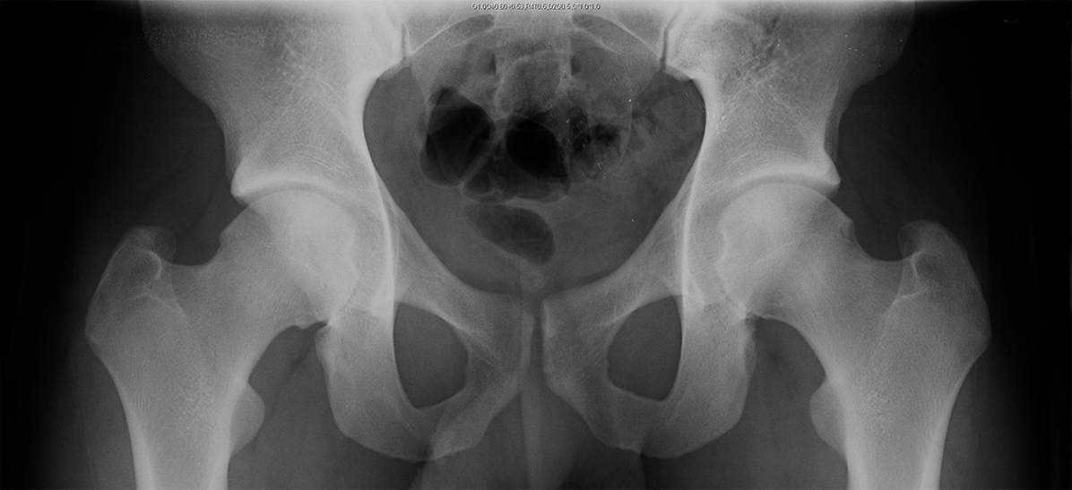
Figure 1AP pelvis radiograph demonstrates bilateral cam lesions.

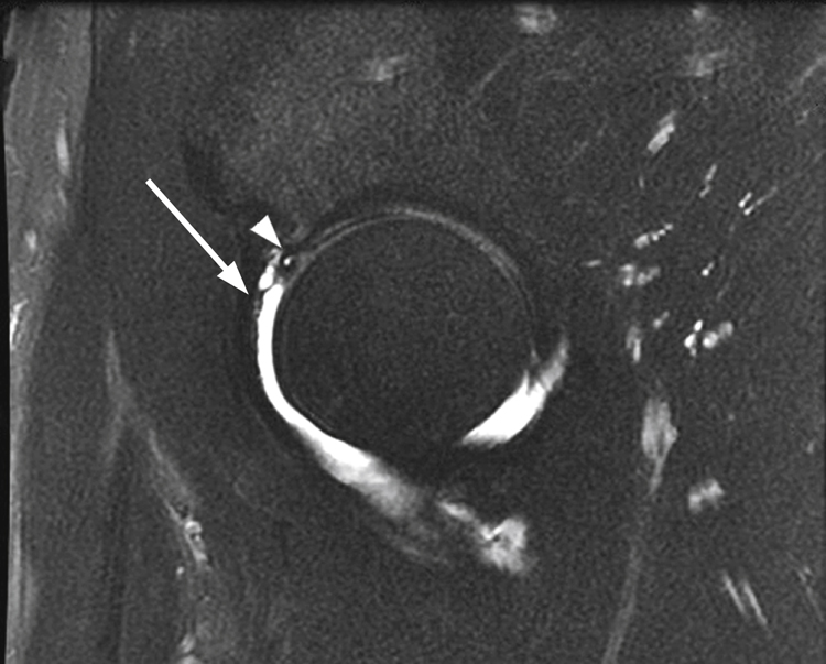
Figure 2Sagittal T2-weighted magnetic resonance arthrogram demonstrates a paralabral cyst (arrow) and a perforation in the anterosuperior labrum (arrowhead).
Standard AP pelvis radiographs
Distance between sacrococcygeal junction and pubic symphysis should be 32 mm in males and 47 mm in females
Lateral center-edge angle (LCEA)—Borderline normal is 20° to 24°; dysplastic is less than 20°
Pincer impingement—LCEA greater than 39°
Tönnis angle—Represents acetabular volume and femoral head coverage
Horizontal line drawn from teardrop to teardrop, and a line tangential to sourcil
Normal is −10° to 10°
>10° indicative of acetabular dysplasia
<−10° indicative of pincer deformity
Dunn view—Obtained at 45° or 90° used to evaluate femoral head sphericity and contour of head-neck junction
Alpha angle characterizes anterior deformity of cam type (>50° for cam)
AP and frog-lateral or cross-table lateral hip radiographs to assess
Acetabular version
Presence/absence of cam lesion (Figure 1)
Herniation pits in femoral neck
Joint space narrowing
Os acetabuli
False-profile view—To measure anterior CEA
CT—Three-dimensional reconstructions help determine bony abnormalities
Magnetic resonance arthrography—Better than MRI at assessing intra-articular soft-tissue pathology (labral tears)
MRI/MRA findings
Paralabral cysts (Figure 2)
Herniation pits at head-neck junction
Os acetabuli
| Video 10.1 Arthroscopic Management of Pincer- and Cam-Type Femoroacetabular Impingement. Christopher M. Larson, MD; Rebecca M. Stone, ATC (8 min) |
Procedure
Room Setup/Patient Positioning

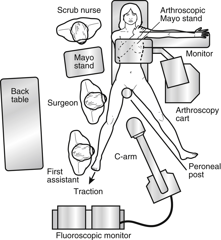
Figure 3Illustration depicts the room setup for hip arthroscopy.

Stay updated, free articles. Join our Telegram channel

Full access? Get Clinical Tree


