Fig. 18.1
Drop vertical jumps. NH not holding a stick (a), RH holding a stick with right hand (b), RHT holding a stick with right hand with a target (c)
18.2.3 Data Collection
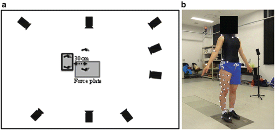
Motion measurements were taken using a 3D optical motion analysis system composed of eight infrared cameras (MotionAnalysis, Kissei Comtec Co., Ltd., Matsumoto, Japan) and a force plate (Kistler Japan Co., Ltd., Tokyo, Japan). Reflective markers were analyzed at a sampling frequency of 240 Hz and ground reaction forces at 2,400 Hz (Fig. 18.2a). The reflective markers were attached to nine points on the skin (the midpoints of and below the left and right anterior superior iliac spine and the left and right superior posterior iliac spine, the right greater trochanter, the medial and lateral epicondyles of the femur, the medial and lateral malleolus, and the second metatarsal head) as well as ten points on the thigh and six points on the calf according to the point cluster method (Andriacchi et al. 1998) (Fig. 18.2b). Three reflective markers were also attached to the heels of the shoes to determine the positions and orientations of foot segments (Fig. 18.2b).


Fig. 18.2
Camera setting (a) and marker position (b)
18.2.4 Kinematic, Kinetic and Data Analysis
The angles of flexion and abduction for the knee joint as well as for the angles of flexion and adduction for the hip joint were calculated from the obtained 3D coordinate data using a joint coordinate system described by Grood and Suntay (1983). Knee joint abduction moments and hip joint adduction moments were also calculated from ground reaction force data using inverse dynamics. Joint moments were calculated as external moments. Each joint moment was normalized by dividing the moment by the product of the subject’s height and weight. The period analyzed was from the point where part of the foot contacted the floor (initial contact: IC) to the point where the tips of the toes left the floor (toe off: TO) based on data obtained from the force plate; this period was designated as the landing phase. The kinematic parameters were peak knee flexion angle, peak knee abduction angle, peak hip flexion angle, and peak hip adduction angle between IC and the 30 % point of the landing phase where the total duration of the landing phase was 100 %. The kinetic parameters were peak knee abduction moment and peak hip adduction moment between IC and the 30 % point of the landing phase. The peak values between IC and the 30 % point of the landing phase were used as parameters because the peak vertical ground reaction force was observed between IC and the 30 % point of the landing phase, in addition to the fact that many reports indicate that real ACL injuries occur near the peak point of maximum ground reaction force seen after IC (Koga et al. 2010; Krosshaug et al. 2007). For statistical analysis, a one-way analysis of variance was used to compare parameters between different jump conditions; significant items were further analyzed utilizing the Bonferroni multiple comparison method. A significance level of <5 % was adopted.
18.3 Results
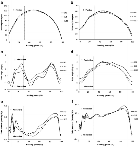
Table 18.1 depicts the mean angles of knee flexion, knee abduction, hip flexion, and hip adduction between IC and the 30 % point of the landing phase, as well as the peak knee flexion angle, the peak knee abduction angle, the peak hip flexion angle, the peak hip adduction angle, the peak knee abduction moment, and the peak hip adduction moment between IC and the 30 % point of the landing phase as measured during all repetitions of NH, RH, and RHT jumps performed by each subject. In Fig. 18.3, time series variations in the mean angles of knee flexion/extension, knee adduction/abduction, hip flexion/extension, and hip adduction/abduction, as well as the mean moments of knee adduction/abduction and hip adduction/abduction for all subjects during each repetition of each jump condition in the landing phase are shown. The peak knee flexion angle between IC and the 30 % point of the landing phase was significantly lower during RHT as compared with RH (p < 0.05). No other significant difference was observed in comparisons between the various parameters of the jump conditions.

Table 18.1
Knee and hip angle and moment during drop vertical jumps (mean ± SD)
NH | RH | RHT | |
|---|---|---|---|
Initial contact | |||
Knee flexion angle (degree) | 26.7 ± 11.6 | 26.4 ± 8.9 | 23.5 ± 6.2 |
Knee abduction angle (degree) | −1.5 ± 3.4 | −1.5 ± 3.7 | −2.3 ± 2.8 |
Hip flexion angle (degree) | 36.8 ± 11.5 | 36.3 ± 8.8 | 34.2 ± 6.6 |
Hip adduction angle (degree) | −5.6 ± 3.2 | −7.8 ± 4.4 | −7.7 ± 4.5 |
Landing phase a | |||
Peak knee flexion angle (degree)b | 113.0 ± 10.6 | 113.5 ± 9.8 | 109.7 ± 10.4 |
Peak knee abduction angle (degree) | −5.1 ± 8.0 | −5.8 ± 7.8 | −6.3 ± 8.0 |
Peak hip flexion angle (degree) | 92.8 ± 7.4 | 91.9 ± 9.3 | 88.6 ± 8.9 |
Peak hip adduction angle (degree) | 2.0 ± 6.2 | 0.4 ± 7.7 | 0.8 ± 7.9 |
Peak knee abduction moment (Nm/(kg*h)) | −0.17 ± 0.14 | −0.15 ± 0.12 | −0.14 ± 0.10 |
Peak hip adduction moment (Nm/(kg*h)) | 0.27 ± 0.10 | 0.30 ± 0.13 | 0.26 ± 0.11 |

Fig. 18.3
Mean knee and hip joint angle and moment during the landing phase: knee joint flexion/extension angle (a), hip joint flexion/extension angle (b), knee joint adduction/abduction angle (c), hip joint adduction/abduction angle (d), knee joint adduction/abduction moment (e), hip joint adduction/abduction moment (f)
18.4 Discussion
This study compared the effect of lacrosse-specific DVJs while holding a stick and while aiming for a target on lower limb kinematics and kinetics in female lacrosse players. The results revealed that the peak knee flexion angle while holding a stick and aiming for a target was significantly smaller than when not aiming for a target. Previous studies have compared lower limb alignment during the DVJ task of female athletes with that of male athletes, and demonstrated that female athletes have greater knee abduction and hip adduction angles, and a smaller knee flexion angle. Many reports state that this dynamic lower limb alignment is a cause of ACL injury in female athletes (Hewett et al. 2006; Renstrom et al. 2008). Furthermore, Decker et al. (2003) reported that during drop landing, female athletes have significantly reduced knee flexion angles as compared to those of male athletes, while Yu et al. (2005) reported that in a stop jump task, the peak female knee flexion angle was significantly smaller. Using data on knee kinematics and kinetics obtained from a DVJ task as baseline values, Hewett et al. (2005) conducted a prospective study comparing an ACL injury group and a non-injury group and demonstrated that the ACL injury group had a significantly greater valgus knee angle than the non-injury group, whereas the peak knee flexion angle as seen from the sagittal plane was significantly smaller (Hewett et al. 2005). Furthermore, Nunley et al. (2003) examined the risk of ACL injury in male and female athletes from an anatomical perspective using X-ray imaging and found that at the same angle of knee flexion, the anterior tibial shear force caused by contraction of the quadriceps femoris muscle was greater in females than males, and thus increased the tensile stress on the ACL.
Stay updated, free articles. Join our Telegram channel

Full access? Get Clinical Tree


