Figure 9.1
Diagram showing the ligaments around the ankle
The distal tibial ossification centre appears between 6 and 24 months of age and the distal fibular ossific centre around 9–24 months of age. The medial malleolus ossifies around 7 years old. The pattern of closure of the distal tibial physis is important:
1.
The centre closes first (this region is known as Kump’s bump or Poland’s hump);
2.
Followed by the medial and;
3.
Finally the lateral side.
This is important in transitional fracture patterns. The process takes around 18 months and is usually complete around the age of 15 years in girls, 2 years later in boys (Fig. 9.2).
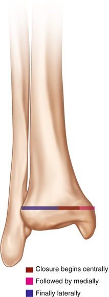
Figure 9.2
Diagram showing the pattern of fusion of the distal tibial physis
9.2 Incidence
Common injuries, accounting for 28–35% of all physeal fractures.
More common in boys than girls.
58% of injuries occur during sports.
Bimodal distribution with small peak in pre-school children and larger peak in adolescents.
9.3 Classification
Distal tibial physeal fractures can be classified anatomically or according to their mechanism of injury.
9.3.1 Anatomical
The Salter-Harris classification can be used as with other physeal fractures. Its advantages are its simplicity and widespread use. It does not, however, describe the forces involved in the injury or differentiate between shearing and crushing mechanisms which carry different prognoses with regards to later growth disturbance.
9.3.2 Mechanism of Injury
The Dias-Tachdjian is the most widely accepted classification based on mechanism of injury and deforming force. Described in 1978, additional categories including axial compression fractures and transitional fractures were added in 1985. It is a modification of the Lauge-Hansen classification. As with the Lauge-Hansen, the first word relates to the position of the foot at the time of injury and second word the deforming force (Fig. 9.3).
Supination-External rotation:
This is the most common mechanism encountered.
Grade 1: Salter-Harris type 2 fracture of the distal tibia; the fracture line extends in a spiral proximally and medially, the distal fragment is displaced posteriorly.
Grade 2: with further external rotation there is an anteroinferior to posterosuperior spiral fracture of the fibula.
Supination-Inversion (Adduction):
Grade 1: distal fibular avulsion (either Salter-Harris type 1 or 2).
Grade 2: further inversion causes a tibial fracture, most commonly a Salter-Harris type 3 or 4. The injury passes through the medial malleolus.
Supination-Plantarflexion.
The tibial epiphysis is forced posteriorly, usually a Salter-Harris type 1 or 2. The fibula remains intact. The tibial fragment may be difficult to appreciate on an AP radiograph. Closed reduction may be difficult as the torn anterior periosteum or the anterior neurovascular bundle may cause soft tissue interposition.
Pronation-Eversion (external rotation).
A Salter-Harris type 1 or 2 fracture of the distal tibia with a transverse fibula fracture. The distal fragments are displaced laterally. Alternatively there may be a Salter-Harris type 3 fracture through the medial malleolus, exiting medial to the midline. This is one of the rare injuries that may be associated with diastasis of the ankle joint in children.
Axial compression.
This causes a Salter-Harris type 5 fracture. Initial radiographs show no abnormality, but follow-up demonstrates growth arrest.
Figure 9.3
(a–d) Summary of the Dias-Tachdjian classification
9.3.3 Transitional Fractures
These fractures occur in the adolescent with a partially closed distal tibial physis.
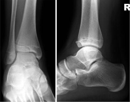
Tillaux fractures (see Fig. 9.4).
Described by Paul Jules Tillaux, a Parisian surgeon (1834–1904).
External rotation injury causes avulsion of the anterolateral distal tibial epiphysis, at the attachment of the anterior inferior tibiofibular ligament.
The injury only occurs when the central and medial parts of the tibial physis have closed but the lateral part remains open.
The typical age is between 13 and 16 years.
The injury is commonly missed.
Figure 9.4
AP and lateral radiographs demonstrating a Tillaux fracture
Triplane fractures.
Described by Lynn in 1972, these occur in the axial, sagittal, and coronal planes.Related posts:

Stay updated, free articles. Join our Telegram channel

Full access? Get Clinical Tree


