Ankle Arthroscopy: Diagnostics, Débridement, and Removal of Loose Bodies
Patient Selection
Indications
TABLE 1
Conditions That Can Be Treated Arthroscopically
| Diagnosis/Indications | Percentage of Cases That Can Be Treated Arthroscopically |
| Ankle arthritis | 25 |
| Loose body | 90 |
| Synovectomy | 90 |
| Acute infection | 80 |
| Lateral impingement | 95-100 |
| Osteochondral defects | 95-100 |
| Anterior osteophytes | 90-95 |
| Stabilization | 25 |
| Ankle arthrodesis | 50 |
| Foreign body | 90-100 |
The most common indications for ankle arthroscopy are listed in Table 1
Anterior impingement
Common with activities requiring forced dorsiflexion
Incidence is up to 45% in football players, 59% in dancers
Physical examination reveals pain on anterior joint line, worse with forced dorsiflexion of the ankle
Arthroscopy removes anterior osteophytes on the tibia or talus
Anterolateral soft-tissue impingement
Involved structures: Any combination of the superior portion of the anterior inferior tibiofibular ligament (AITFL), the distal portion of the AITFL, along the anterior talofibular ligament (ATFL), lateral gutter near talar dome
Physical examination reveals ankle pain worsened by forced dorsiflexion, pain in the lateral gutter
Contraindications
Infection
Neuropathy
Complex regional pain syndrome
Psychiatric disorder
Preoperative Imaging
Radiography

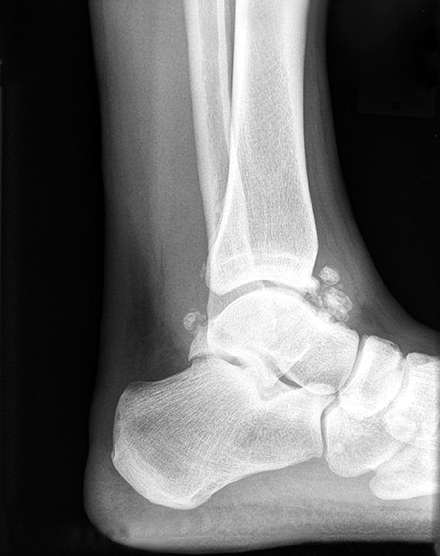
Figure 1A lateral radiograph of the ankle reveals an. anterior tibial spur.
Lateral ankle view (Figure 1) may show anterior osteophytes on tibia, talus with dorsal spur, or signs of dorsal degeneration
Dorsiflexed lateral ankle view may show impingement
Magnetic Resonance Imaging

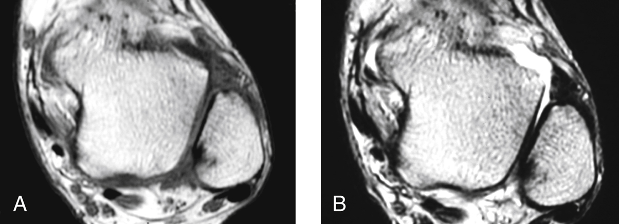
Figure 2Axial T1-weighted (A) and fast spin-echo T2-weighted (B) MRIs of the ankle of a 39-year-old man with anterolateral ankle impingement. Thickening and scarring of the distal fascicle is evident in these images taken 3 mo after an anterior inferior tibiofibular ligament injury.
Not indicated in all cases of anterior impingement
Common findings—Cartilage thinning, soft-tissue swelling, osteophyte
Injury to the AITFL or ATFL is best visualized on short tau inversion recovery and sagittal T1-weighted images (Figure 2)
MRI is 79% accurate and 84% sensitive in diagnosing anterior soft-tissue impingement
Ultrasonography
Can identify synovial lesions in anterior lateral gutter and ligament injuries
Stay updated, free articles. Join our Telegram channel

Full access? Get Clinical Tree


