End-Stage Ankle Arthritis
Arthrodiastasis, Supramalleolar Osteotomy, or Arthrodesis?
Keywords
• Ankle • Arthritis • Arthrodiastasis • Supramalleolar osteotomy • Arthrodesis
Arthritis
Osteoarthritis is a degenerative disease of joints characterized by formation of osteophytes, subchondral sclerosis, subchondral cysts, loose bodies, and joint space narrowing.1–3 It affects approximately 15% of the world’s population, of which 1% is suffering with osteoarthritis of the ankle.2 In the United States, arthritis is the leading cause of disability. About 21 million people reported having arthritis, and subsequent limitation of their work-related function has been found in 1 out of 3 of these people.4 Daily function is significantly affected compared with the general population.5 According to Glazebrook and colleagues,3 end-stage ankle arthritis has a severe impact on pain, health-related quality of life, and function that is at least as severe as patients with end-stage hip arthritis. In general, patients with end-stage ankle arthritis experience greater emotional and mental distress than those who are experiencing end-stage hip arthritis.
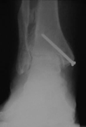
The causes of osteoarthritis can be divided into 3 categories: primary, secondary, and posttraumatic. Primary osteoarthritis is idiopathic in nature with no obvious underlying abnormalities occurring 50% of the time, whereas secondary osteoarthritis occurs in patients with underlying conditions such as rheumatoid arthritis, hemarthroses, hemophilia, and postinfectious processes. Although primary osteoarthritis is the most common cause of hip and knee problems, the same is not observed in the ankle.3,5–10 Primary osteoarthritis of the ankle affects older populations of patients. The primary group also has less pain and increased range of motion compared with secondary and posttraumatic osteoarthritis groups.2,6,11 Valderrabano and colleagues2 evaluated 406 ankles with symptomatic end-stage osteoarthritis. In their study, posttraumatic osteoarthritis of the ankle was seen in 78% of cases, secondary osteoarthritis in 19% of cases, and primary osteoarthritis in 9% of cases. Similar results were found by Saltzman and colleagues,6 evaluating 639 ankles with 70% of cases occurring secondary to trauma of the ankle joint. Malleolar ankle fractures, ligamentous injuries causing ankle instability, pilon tibial fractures, tibial shaft fractures, talus fractures, osteochondritis dissecans, and severe combined fractures were the main causes of posttraumatic osteoarthritis of the ankle seen in both studies (Fig. 1).
Nonoperative care
Conservative treatments are limited for symptomatic end-stage ankle arthritis. Most therapies provide short-term improvement of symptoms and should be exhausted before consideration of surgical treatment options. Nonoperative, conservative treatment options include a combination of medications, injections, modification of activities, prescription of custom orthotic devices, and bracing.12–14
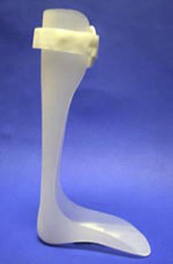
Nonsteroidal antiinflammatory drugs (NSAIDs) may help relieve pain of arthritic ankle joints. They should be given only short term and closely monitored for side effects. Altered kidney function tests as well as bleeding tendencies are the most common side effects associated with NSAIDs. A combination of corticosteroid-anesthetic intra-articular injection can be given to decrease joint pain and inflammation. Varied results have been reported for the duration of beneficial effects of the injection. Side effects are uncommon but skin depigmentation and infections may be seen. Modification of activities may be beneficial. Patients’ pain may be more manageable with changes in occupation to a sedentary job as well as a decrease in vigorous activities such as sports. Pain and inflammation can also be managed with bracing and change in shoe gear. Rocker-bottom sole, solid ankle cushion heel (SACH), lace-up ankle support braces, ankle-foot orthosis, and weight-bearing fiberglass or plaster cast can decrease inflammation and pain by restricting motion of the ankle joint. Patella tendon–bearing (PTB) braces have also been used with some success for treatment of ankle arthritis because they reduce pain and discomfort of the affected extremity by decreasing axial load (Fig. 2).12–14
Arthrodiastasis
The term arthrodiastasis comes from the Greek words arthro (joint), dia (through), and tasis (to stretch out). Distraction of the ankle joint has been used as an alternative to arthrodesis or arthroplasty. This procedure is advocated to reduce pain and increase motion of an arthritic joint without sacrificing the joint. It is indicated in younger patients with good bone stock and painful ankle joints who are not willing to have an ankle arthrodesis.15,16
The technique was first described by Judet in 1978 for treatment of osteoarthritis of the hip.17 It was not until 1995 that van Valburg and colleagues17 reported on the use of an arthrodiastasis technique for treatment of severe posttraumatic arthritis of the ankle joint. The Ilizarov external fixator was used for ankle distraction in 11 patients in combination with measurement of intra-articular hydrostatic pressure. The Ilizarov external fixator was applied for 3 months and the ankle joint was distracted 5 mm. Patients were able to be fully weight bearing just days after surgery. At 3 months, the fixator was removed and patients were transitioned into a cam boot. Clinical improvement of pain and mobility was observed at mean follow-up of 20 months and an increase in joint space was also noted on weight-bearing radiographs. During loading, the researchers observed an increase in intra-articular pressures of the distracted ankles.
After distraction of a joint, theoretically the cartilage has the potential to repair itself. It is thought that mechanical off-loading can prevent further damage to the articular cartilage. Once the joint is off-loaded, the chondrocyte repair process may begin with fluctuation in intra-articular hydrostatic pressure during weight bearing with the external fixator. Chondrocytes are able to repair by the cyclic changes in intra-articular fluid pressure within the joint.15,18,19
In 2002, Marijnissen and colleagues20 published a large multicenter prospective study of 57 patients with a mean age of 44 years who underwent ankle distraction and ankle arthroscopy when necessary. Patients were followed on average of 2.8 years. Eleven of the 57 patients were excluded from the study because of short follow-up of less than 1 year and 13 patients withdrew from the study because of recurrent pain and required further treatment. Significant clinical improvement was seen in 38 patients at 1-year follow-up. More importantly, significant functional and clinical improvement was seen compared with the results at 1 year. A randomized study on 17 patients was also performed by the investigators. They evaluated 9 patients with ankle distraction with arthroscopic debridement as necessary compared with 8 patients with arthroscopic debridement alone. The results from the ankle distraction group were similar to their prospective study. In the debridement group, significantly less profound outcomes were observed and 3 of the 8 patients did not reach 1-year follow-up. The failures underwent joint distraction with satisfactory results. In this large multicenter study, significant improvement was observed using joint distraction.
Short-term results of joint distraction have proved to be satisfactory.
Ploegmakers and colleagues21 performed a multicenter retrospective analysis of 27 patients with posttraumatic osteoarthritis. All patients were treated with Ilizarov ankle distraction. Of the 27 patients, 2 could not be traced and 3 patients incorrectly completed the questionnaire and could not be included in the study. Data was evaluated for these 22 patients with a mean age of 37 years and at least a 7-year follow-up. Six of these patients had remaining persistent pain and went on to arthrodesis. Sixteen patients were evaluated on the basis of pain, function, clinical status, and mobility at a mean 10-year follow-up. Sixteen of the 2-2, or 73%, of patients had significant improvement in all clinical parameters evaluated.
The data from multiple studies with large patient populations as well as long-term results show improvement of symptoms and function following ankle joint distraction in patients with severe posttraumatic osteoarthritis. Ankle joint distraction provides a viable joint-sparing treatment of ankle osteoarthritis. Most studies suggest that younger patients benefit more from ankle distraction, although Tellisi and colleagues15 evaluated age as a predictor of results and showed that patients older than 60 years had more improvement. Even though relief or improvement of symptoms may be temporary, more definitive treatment, such as ankle arthrodesis, can be considered at a later date.
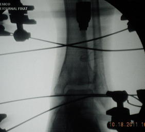
It is the experience of the authors that this provides a reasonable option for patients with end-stage ankle arthritis, in particular for younger patients. The authors suggest that this procedure be reserved for the right patient, and suggest that the patient be fully engaged in preoperative detailed demonstration and explanation of the procedure. In review of the authors experience, we think the condition surrounding joints contributes to the success or failure of the procedure (Fig. 3).
Ankle arthrodesis
Ankle arthrodesis is a well-documented surgical treatment of end-stage ankle arthritis. It has been a preferred treatment of ankle arthritis because of its predictable outcomes. In 1879, Albert was the first to described ankle arthrodesis to treat paralytic ankle equinus.14 More than 30 different techniques to improve the results of the procedure have been described since that time. Ankle arthrodesis is indicated when patients experience persistent pain secondary to the deformity that limits their daily function and after all conservative treatment options have failed.13,14,22,23 Although still considered the gold standard, ankle arthrodesis for treatment of painful end-stage arthritis, clinicians must be aware of the biomechanical effects on the lower extremity and surrounding joints.
In earlier studies, surgeons experienced high nonunion rates and a higher need for revision surgery. Recent literature reports higher rates of fusion, although variability of fusion rates do exist. Studies report successful union rates of 81% to 99%.22,24–27 High rates of nonunion have been associated with use of the external compression clamp that was popularized by Charley.22,26,27 In contrast, in ankle arthrodesis, internal fixation has been associated with higher rates of union.24,27–30 Morgan and colleagues28 reviewed 101 ankle joint fusions with an average follow-up for patients of 10 years. An anterolateral surgical approach was used to gain access to the ankle joint and arthrodesis was achieved with screw fixation. A 95% fusion success rate was reported, which can be attributed to their emphasis on preparation of the joint to achieve bone-on-bone contact and use of internal fixation. Zwipp and colleagues24 reported a fusion rate of 99% in 93 out of 94 patients using a 4-screw technique. Using an anterior fusion plate, Rowan and colleagues27 achieved a 92% fusion rate in 31 out of 34 patients.
Following ankle arthrodesis, patients notice significant decrease in steps per minute, in addition to decreased stride length, but do not have a significant amount of pain compared with the control group. No significant difference in range of motion in the sagittal plane of the pelvis or the knee joint was seen but fusion of the ankle showed significant decrease in range of motion of the hindfoot and forefoot in all planes (sagittal, transverse, and frontal).31 Buck and colleagues32 studied the importance of position of ankle fusion and its affects on patterns of motion of the hindfoot and effect of different ground conditions. The recommended that the optimal position of the ankle joint is neutral flexion, 0° to 5° of valgus of hindfoot angulation and 5° to 10° of external rotation of the foot. A dorsiflexed position is better tolerated than a plantarflexed position of ankle fusion because it decreases sagittal plane motion of the foot and also causes genu recurvatum, producing an abnormal gait that is exaggerated with different ground conditions. Increased extension of the knee is also caused by anterior position of the talus on the tibia and during ambulation uphill. Varus hindfoot position produces a supinated foot type causing locking of the midtarsal joint, whereas arthrodesis in a slight valgus position allows greater motion in the foot. In the stance phase of gait, internal rotation of the foot decreases hindfoot motion and external positioning is indicated to decrease medial collateral ligament stress during toe-off.
Most patients who undergo ankle arthrodesis are satisfied with their results and would go through surgery again in the same circumstances.25,28,30–33 However, they have long-term functional limitations secondary to pain in adjacent joints.34 Early postoperative results show no significant changes in adjacent joints, but in one long-term study35 and another by Coester and colleagues34 there were significant arthritic changes in adjacent ipsilateral joints compared with the contralateral extremity. Patients had increased osteoarthritic changes in subtalar, talonavicular, and calcaneocuboid joints. In a long-term follow-up study on quality of life by Fuchs and colleagues,33 17 patients were followed up for at least for 20 years after ankle arthrodesis. Charnley compression clamps were used in 14 patients as an external fixator for ankle arthrodesis. Only half of the patients had minor restrictions of activities of daily living. Sixteen of the 17 patients were still working and 44% returned to their preinjured occupations, with others performing lighter duties. Similar results were found by Buck and colleagues.32 Patients had increased physical limitations, emotional disturbance, and pain compared with the age-matched normal population. Significant correlation between functional outcome and radiographic osteoarthritic changes were seen in subtalar joints but not in midtarsal joints. A larger study of 107 subjects was performed by Slobogean and colleagues.36 Their prospective study evaluated patients with ankle arthrodesis and ankle arthroplasty and their health state values using an SF-36 generic health-related quality of life instrument. The SF-36 uses 11 items to create 6 dimensions (SF-6D), namely physical function, role limitation, social functioning, bodily pain, mental health, and vitality. Patients were evaluated at baseline and at 1 year. They found no statistical difference in results at baseline or 1 year between the ankle arthrodesis or arthroplasty groups. Significant improvement in SF-6D scores were seen between baseline and 1-year follow-up of ankle arthrodesis and ankle arthroplasty groups. At 1-year follow-up, patients’ SF-6D results approached age-matched and gender-matched US population norms.
Jung and colleagues37 evaluated 12 cadaver limbs with an average age of specimen of 68 years (range 52–88 years). A 700-N load was tested on all cadaver specimens. The researchers measured joint contact pressures, peak pressure, and contact area in the talonavicular, subtalar, and calcaneocuboid joints before and after immobilization at neutral ankle axial loading and at tibiopedal dorsiflexion at different angles. Evaluation of different angles was meant to simulate late stance phase of the gait cycle. The results showed that there was significant increase in contact and peak pressures in talonavicular and calcaneocuboid joints between intact and fused ankles at different degrees of dorsiflexion. Comparison of the subtalar joint in intact and fused ankles showed no significant difference in contact or peak pressures but had an increase in contact surface area. Similar results were recorded by Suckel and colleagues38 in 8 cadaver specimens. Further, an increase in peak pressures were seen at the talonavicular joints. These results suggest that increase in peak pressures at the talonavicular joint may lead to cartilage degeneration and long-term pain along the medial column.
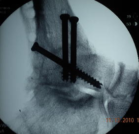
Many approaches have been described. They consist of anterior, anterior-lateral, medial, lateral, transmalleolar, and posterior. It is the authors experience that the anterior and posterior approach allow the best ease of correction, especially with a frontal plane deformity. Each approach has its own benefit and downside. The posterior approach is favored when there is soft tissue compromise because the soft tissue envelope is thicker and rich in vascularity because of a low-lying flexor hallucis muscle below. Joint preparation can be performed as either curettage, joint resection, burring, or fish scaling. Each of these techniques has its own advantages and disadvantages too. It is the authors’ experience that the curettage technique allows the least amount of shortening, provides excellent contour and inherent stability, with excellent bone-to-bone contact, therefore it is the technique of choice of the authors. Fixation options consists of internal fixation and external fixation. The internal fixation for the tibial-talar joint can be a choice of screws, staples, and plates. The authors’ first choice is to use large cancellous screws combined with a locking plate and an onlay graft from the fibula as a biologic fixation. The authors highly recommend leaving the fibula intact because this allows patients and the surgeon the option of performing a takedown in the future. This method allows for the possibility of an ankle arthrodesis to be converted to an ankle replacement if needed (Figs. 4–9).


Stay updated, free articles. Join our Telegram channel

Full access? Get Clinical Tree


