CHAPTER 7 ANKLE AND FOOT
A LIGAMENT STRESS TESTS
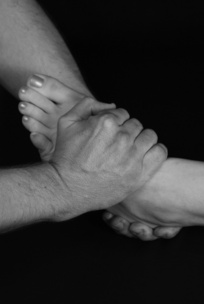
Anterior talofibular ligament stress test
Technique
Action
The calcaneum is tilted into a plantarflexed position. The upper hand then gradually adds further plantarflexion and inversion (see Fig 7.1).
Clinical context
The ATFL is the most important lateral stabilizer of the ankle and the most frequently injured (Tohyama et al 1995, Trojian & McKeag 1998, Wolfe et al 2001). With the foot in a neutral position, the fibula–ATFL angle is around 90° (i.e. the ligament runs approximately parallel to the sole of the foot) but plantarflexion brings it increasingly parallel to the long axis of the fibula where it functions as the main collateral ligament (Bahr et al 1997). In increasing degrees of plantarflexion and inversion, strain of the ATFL increases, more so than the calcaneofibular ligament (CFL), thereby rendering the ATFL most vulnerable in this position (Bahr et al 1997, Colville et al 1990). The posterior talofibular ligament is the strongest of the lateral complex and is only injured in a severe inversion sprain (Wolfe et al 2001).
While careful physical examination has been shown to be a valuable tool in the detection of ankle fracture (see clinical tip; Stiell et al 1994), accurate evaluation of ligament injury is more difficult (Fujii et al 2000, Van Dijk et al 1996). Isolated tears do not tend to produce significant instability and pain is usually the dominant finding on stress testing. If the trauma is more severe and accompanied by swelling and bruising, instability testing should be performed (i.e. drawer test, p. 250, and talar tilt test, p. 248) with the index of suspicion high of a double rupture of the ATFL and the CFL, if positive (Bahr et al 1997).
Clinical tip
Leaving the pain and swelling to settle for a few days has been shown to be advantageous in improving the diagnostic accuracy of ligament injury at the ankle (Van Dijk et al 1996).
The widely accepted Ottowa rules for radiographic examination post ankle trauma have been found to produce a very low rate of false negatives and a sensitivity of 100%, reducing the number of ankle X-rays by about 35% (Stiell et al 1992). A subsequent larger trial confirmed these findings (Stiell et al 1994) which led to effective implementation at multiple centres (Stiell et al 1995). The Ottowa rules (Duckworth et al 2009) state that:
An ankle X-ray is only required if there is:
A foot X-ray is required if there is:
| EXPERT OPINION | COMMENTS |
|---|---|
 | ATFL stress test |
| An extremely useful test to preferentially stress the ATFL but more valuable once the acute stage has passed and sufficient plantarflexion is available. This can also be used as a global lateral ligament stress test. |
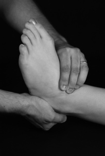
Calcaneofibular ligament stress test
Technique
Clinician position
The calcaneum is cupped by one hand (right foot/left hand and vice versa) while the other hand wraps over the dorsum of the foot, the fingers positioned over the lateral talar dome and the thumb supporting the sole of the foot.
Clinical context
This test is exactly the same as the manoeuvre described at the talar tilt test but with the emphasis on the detection of minor ligamentous injury rather than ankle instability (see talar tilt, p. 248). With the foot in the neutral position, the CFL forms a posterior angle of about 130° with the fibula, but with the foot in dorsiflexion the ligament becomes parallel to the axis of the fibula, thereby functioning as a collateral ligament. As a result, the ligament is under most stress in dorsiflexion and inversion while its lateral companion, the anterior talofibular ligament (ATFL), provides little restraint in this position (Bahr et al 1997, Colville et al 1990). With the reverse occurring in plantarflexion and inversion, it is clear that the ATFL and CFL function together in all positions of ankle flexion to provide lateral ankle stability (Colville et al 1990).
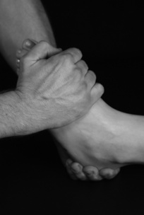
Calcaneocuboid ligament stress test
Technique
Action
The calcaneum is fixed in a neutral position while the other hand applies a combined movement of adduction and inversion of the forefoot (see Fig. 7.3).
Clinical context
The CCL is composed of the dorsal CCL ligament (a thickening of the fibrous capsule on the dorsal surface of the joint) and the CCL component of the bifurcate ligament (Standring 2005). Compared to the prevalence of anterior talofibular ligament (ATFL) injury, the CCL is much less commonly involved in ankle sprains. It can either be injured in isolation, where the forefoot is exposed to forced inversion and adduction while the calcaneus is relatively fixed and stable, or as a combined lesion resulting from gross lateral strain. In ankle sprains, the ATFL, calcaneofibular ligament and the CCL can all be involved and the ligaments should always be tested for pain and laxity (i.e. ATFL stress test, p. 236; drawer test, p. 250; talar tilt test, p. 248). If the CCL is involved as part of a combined sprain, the ATFL is most likely to be its injured partner.
Clinical tip
The calcaneocuboid joint line can be found by placing the side of the thumb up against the base of the fifth metatarsal where the joint will be approximately in line with the midpoint of the thumb pad. It is important to ensure that the hand is positioned just distal to the joint line in order to localize stress on the ligament effectively.
| EXPERT OPINION | COMMENTS |
|---|---|
 | CCL stress test |
| The CCL is often missed as a component of a lateral complex sprain and this test permits more specific localisation. |
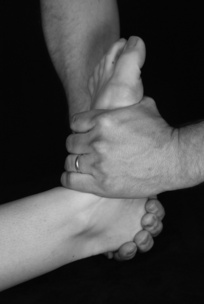
Medial collateral ligament stress test
Technique
Action
The calcaneum is tilted into a valgus position while the upper hand gradually adds eversion in a degree of dorsiflexion (see Fig. 7.4).
Clinical context
The medial ligament is a very strong fan-shaped structure (composed of the tibionavicular, tibiocalcaneal, anterior and posterior tibiotalar ligaments) which limits eversion of the ankle and lateral displacement of the talus. It is made up of superficial bands which are mainly vertically orientated, limiting rear foot eversion, and deeper fibres, more transverse in direction, which limit abduction/external rotation of the talus (Placzek & Boyce 2006). Its strength is demonstrated by the fact that the malleolus often fractures before the ligament ruptures – 75% of ankle fractures occur on the medial side. Conversely, medial ligament injury represents only 10% of ankle sprains (Trojian & McKeag 1998) and this is attributed to the enhanced medial stability afforded by the mortise of the ankle, the articulation between the medial malleolus and talus and the anterior tibiofibular ligament, all of which make injury much less likely than on the lateral side (Wolfe et al 2001).
Injury to the medial ligament will be evident by the mechanism of injury (excessive eversion with the foot in a neutral or slightly dorsiflexed position), local tenderness, swelling and a positive medial ligament test. If significant trauma has occurred other injuries should also be considered such as a syndesmosis injury (see external rotation stress test, p. 244) or fracture, i.e. Maisonneuve fracture (proximal fibula), distal fibular fracture or avulsion fracture of the medial malleolus (Trojian & McKeag 1998) requiring further evaluation (see Ottowa rules, p. 238) and possible surgical intervention.
Clinical tip
The medial ligament is also vulnerable to chronic strain resulting from poor foot biomechanics. A valgus rear foot and associated pronation of the forefoot will produce medial forces that, if left uncorrected, may lead to chronic strain on both the medial and spring ligaments. The stress test may well reveal some discomfort at the extreme of the movement and, if very established, a small degree of laxity may be evident. Because this is usually a bilateral problem, making a comparison with the ‘normal’ side is not always possible.
| EXPERT OPINION | COMMENTS |
|---|---|
 | MCL stress test |
| Primarily evaluates the superficial fibres, which are more commonly involved in biomechanical overloading and minor injury. |


