Fig. 1.1
Anatomy of patella. (a) Anterior view. (b) Lateral view. 1. Base. 2. Apex. 3. Femur. 4. Quadriceps tendon.5. Patellar tendon. 6. Tibia. 7. Fibula. 8. Posterior medial facet. 9. Posterior lateral facet
The posterior surface of the patella is articular area and has three facets:
One concave lateral facet (representing 2/3 of the articular surface)
One more convex medial facet (representing 1/3 of the articular surface)
One facet located on the medial border of the patella (“odd facet”)
The medial and lateral facets are separated by the vertical patellar ridge which conforms to the trochlear groove.
Articular congruence between the patella and the femur is imperfect and varies according to the position of the knee:
In the early degrees of knee flexion, contact is made exclusively on the distal part of the articular surface.
As flexion increases, the contact area moves proximally. The maximum surface contact is at 45° of flexion.
At 90° of flexion, the contact is located in the proximal portion of the patellar articular surface.
The “odd facet” does not articulate except in maximum flexion, starting at 135°.
The Distal Femur [12–19]
The femoral condyles constitute the articular surface of the femur and are asymmetrical:
The lateral condyle is wider and shorter and is positioned more sagittally. The radius of curvature decreases significantly from front to back (Fig. 1.2).
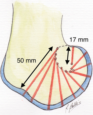
Fig. 1.2
Curvature of femoral condyle
The medial condyle has a long axis oriented approximately 22° with respect to the sagittal axis. Its radius of curvature is more regular than the lateral condyle.
At the anterior surface, the two condyles are separated by a depression: the femoral trochlea. The trochlear groove is the deepest part of femoral trochlea. It articulates with the posterior articular surface of the patella. The lateral side of the trochlea is more prominent than the medial one.
The intercondylar notch separates the two femoral condyles at their distal and posterior part (Fig. 1.3).
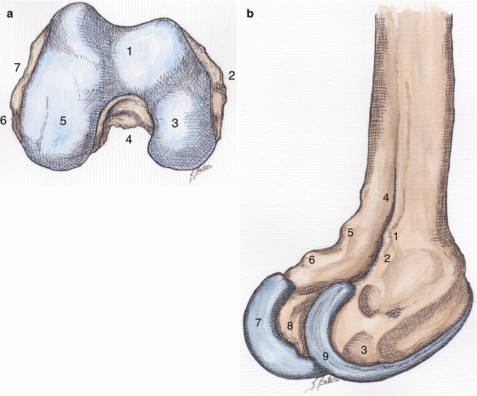
Fig. 1.3
Anatomy of distal part of the femur. (a) Inferior view. 1. Patellar. 2. Medial epicondyle. 3. Medial condyle. 4. Intercondylar notch. 5. Lateral condyle. 6. Insertion area of popliteal tendon. 7. Lateral epicondyle. (b) Posterolateral view. 1. Supracondylar lateral tuberosity. 2. Footprint of the lateral collateral ligament. 3. Insertion area of popliteal tendon. 4. Popliteal notch. 5. Adductor tubercle. 6. Supracondylar medial tuberosity. 7. Medial condyle. 8. Intercondylar notch. 9. Lateral condyle
The anterior cruciate ligament (ACL) originates from its lateral wall, and the posterior cruciate ligament (PCL) originates from the medial wall.
The lateral epicondyle is a prominence of the lateral aspect of the knee, receiving the insertion of the lateral collateral ligament. It is separated from the distal articular surface by a depression, where the popliteal tendon originates.
The medial epicondyle is a prominence of the medial aspect of the knee, receiving the origin of the medial collateral ligament. It is located anteriorly and distally relative to the adductor tubercle.
There exists between the epicondylar axis and the posterior condylar line a mean external rotation of 3.5° in men and 1° in women.
In the mediolateral aspect, women tend to have a narrower femur relative to men.
The Proximal Tibia [20]
The medial and lateral part of the tibial plateau conforms to the femoral condyles and to the lower surfaces of the menisci:
The lateral tibial plateau is smaller and more rounded. Its articular surface is convex.
The medial tibial plateau is larger and more oval. Its articular surface is concave.
Their major axis is oblique forward and laterally, and both of them have a posterior slope of about 10° relative to the tibial diaphysis.
The tibial spine is a prominence of the middle part of the tibia. The pre-spinal surface receives insertions of several structures. From anterior to posterior, these are the anterior horn of the medial meniscus, the anterior cruciate ligament, and the anterior horn of the lateral meniscus.
The retro-spinal surface is found posterior to the spine. From anterior to posterior, this receives the insertions of the posterior horn of the lateral meniscus, the posterior horn of the medial meniscus, and the posterior cruciate ligament at the level of the tibial margin.
Between these two surfaces are located the medial and lateral intercondylar tubercles, separated by the intertubercular fossa. The medial tubercle is higher and more anterior than the lateral tubercle.
The articular congruence between the tibial plateau and femoral condyles is increased by the presence of the tibial spine and intercondylar tubercles, in addition to the menisci.
Two bony prominences are found on the anterior surface of the tibial epiphysis:
The tibial tuberosity, located below the pre-spinal surface, which is the insertion zone of the patellar tendon
Gerdy’s tubercle, located 2–3 cm lateral to the tibial tuberosity, which receives the distal insertion of the fascia lata (iliotibial band)
Proximal Tibiofibular Joint
The proximal part of the fibula is composed of the head, which articulates with the tibia with its anteromedial part. At its superior part are inserted the distal part of the lateral collateral ligament, the tendon of biceps femoris, the fabellofibular ligament, and the arcuate ligament.
Below the head is the neck, the narrow portion of the fibula in contact with the common peroneal nerve (Fig. 1.4).
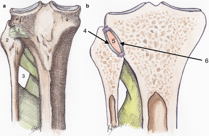
Fig. 1.4
Proximal tibiofibular joint. (a) Anterior view. (b) Frontal cut. 1. Anterior tibiofibular ligament. 2. Fibular head. 3. Crossing of tibial anterior artery. 4. Articular surface of the fibular head. 5. Synovial cavity. 6. Articular surface of the tibia
The proximal tibiofibular joint has a synovial membrane and an independent joint capsule from the knee joint.
Fabella
The fabella is a sesamoid bone of the knee, located in its dorsal surface.
It is incorporated within the oblique popliteal ligament (Fig. 1.5).
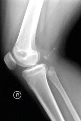
Fig. 1.5
Fabella, radiological view (arrow)
1.1.2 Articular Cartilage [21]
Articular cartilage (or hyaline cartilage) covers the bone articular surfaces and allows sliding between them.
It is a tissue without innervation and without vascularization. It obtains nutrition by diffusion from the synovial fluid and the subchondral bone.
The normal articular cartilage in adults has a white surface, slightly translucent, smooth, and shiny. It has a certain flexibility and deformability.
The thickness varies depending on the joints. The knee cartilage is very thick: 6–7 mm at the patellofemoral joint, 5–6 mm at the femoral-tibial compartment.
The cartilage is composed of:
Chondrocytes, a mature cell population with very little activity and sparsely distributed, synthesizing the extracellular matrix and regulating cartilage homeostasis
Extracellular matrix rich in water (65–80 % wet weight of cartilage), type II collagen (providing cartilage its compressive strength), and proteoglycans
It has multiple layers, which differ from each other on biochemical (proteoglycan content) and anatomical (complexion of collagen fibers) criteria.
Articular cartilage enables the sliding of the bone surfaces, thanks to an extremely low coefficient of friction. It is resistant, elastic, and lubricated by the synovial fluid. It acts as a shock absorber.
1.1.3 Menisci [12, 22–29]
The menisci are two crescent-shaped fibrocartilaginous structures.
Their roles are multiple:
Improving joint congruence between the femoral condyles and tibial plateaus.
Transmission of force and absorption of shocks, due to their viscoelastic properties.
Proprioception of the knee joint, due to their rich innervation.
The menisci are composed of:
An extracellular matrix consisting primarily of type I collagen (70 %), glycosaminoglycans, glycoproteins, and elastin fiber
Fibroblast and fibrocartilaginous cells scattered within the collagen matrix
Collagen fibers are mainly arranged circumferentially, crossed by radial fibers, improving meniscal strength and rigidity (Fig. 1.6).
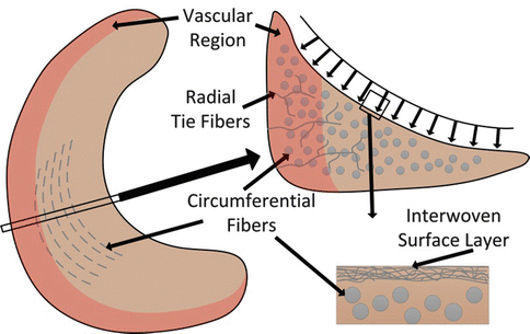
Fig. 1.6
Anatomical structure of the meniscus, organization of collagen fibers
Only the peripheral third of the meniscus is vascularized; the central portion remains avascular.
The menisci have a triangular cross-sectional shape, accepting the convex femoral condyle superiorly and the flatter peripheral tibial plateau inferiorly.
Each meniscus covers about 2/3 of each peripheral tibial plateau.
Their peripheral edge is convex, thick, and attached to the joint capsule, while the central edge is thin and free within the joint.
They consist of three segments: anterior horn, posterior horn, and body.
The posterior horn of the medial meniscus is significantly larger than the anterior horn, while at the lateral meniscus, the horns have identical sizes.
1.1.3.1 Medial Meniscus
The medial meniscus is generally semicircular and measures approximately 3.5 cm.
Its posterior horn is attached just in front of the PCL, while its anterior horn is attached in front of the ACL. The transverse inter-meniscal ligament connects the anterior horns of the medial and lateral menisci.
The peripheral part of the meniscus is attached to the joint capsule and reinforced by the meniscofemoral ligament (a layer of the deep medial collateral ligament) and the meniscotibial (or coronary) ligament to the peripheral tibial margin.
The posteromedial segment of the meniscus receives a portion of fibers of the semimembranosus tendon.
The large posterior horn of the medial meniscus also controls internal tibial translation and acts as a secondary restraint to anterior tibial translation.
1.1.3.2 Lateral Meniscus
The lateral meniscus is almost circular and covers a larger portion of the lateral tibial plateau.
Its posterior horn is attached just in front of the posterior horn of the medial meniscus, while its anterior horn is attached behind the ACL.
Posteriorly, the lateral meniscus is also connected to the medial side of the lateral condyle through meniscofemoral ligaments intersecting the posterior cruciate ligament (Humphrey ligament anteriorly and Wrisberg ligament posteriorly).
The peripheral part of the meniscus is attached loosely to the joint capsule, interrupted at its middle part by popliteal hiatus, which gives passage to the tendon of the popliteal muscle.
Some fibers of the popliteal tendon are inserted in posterior superior edge of the meniscus.
The lateral meniscus is much more mobile, with the ability to move 1 cm anteroposteriorly with knee flexion and lateral femoral condylar rollback.
1.1.4 Capsule and Synovial Cavity [21, 30]
The articular capsule is a fibrous membrane of varying thickness around the knee joint.
It is thin at the anterior part of the knee. It inserts on the periphery of articular part of the patella and on the lateral edges of the patellar tendon.
Proximally, it inserts to the anterior surface of the cortex of the femur three or four fingerbreadths above the patella. It joins the base of the patella on the deep surface of the quadriceps.
Distally, it is inserted around the margin of the tibial plateau and on the periphery of the menisci, except at the popliteal tendon level where it bends to form the popliteal hiatus (popliteus tendon is therefore an intra-articular structure).
Medially and laterally, it is inserted on the periphery of the femoral condyles, about 10 mm from condylar articular surface.
At the posterior part of the knee, the capsule is very thick with vertical fibers making the posterior condylar folds. It goes into the intercondylar notch in contact with PCL, which therefore remains intra-articular. Condylar hulls are reinforced by numerous ligaments derived from semimembranosus and popliteal tendons.
The synovium is a translucent pink tissue that is composed of a specific cell population – synoviocytes.
There are two types of synoviocytes: those having a macrophage function and those having a role for the synthesis of synovial fluid.
The synovial membrane covers the deep surface of the capsule, the suprapatellar bursa located in the quadricipital recess, as well as infrapatellar fat pad located deep to the patellar ligament (“Hoffa’s fat pad”).
It is also found in contact with cruciate ligaments and popliteal tendon (these structures are intra-articular but extra-synovial) (Fig. 1.7).
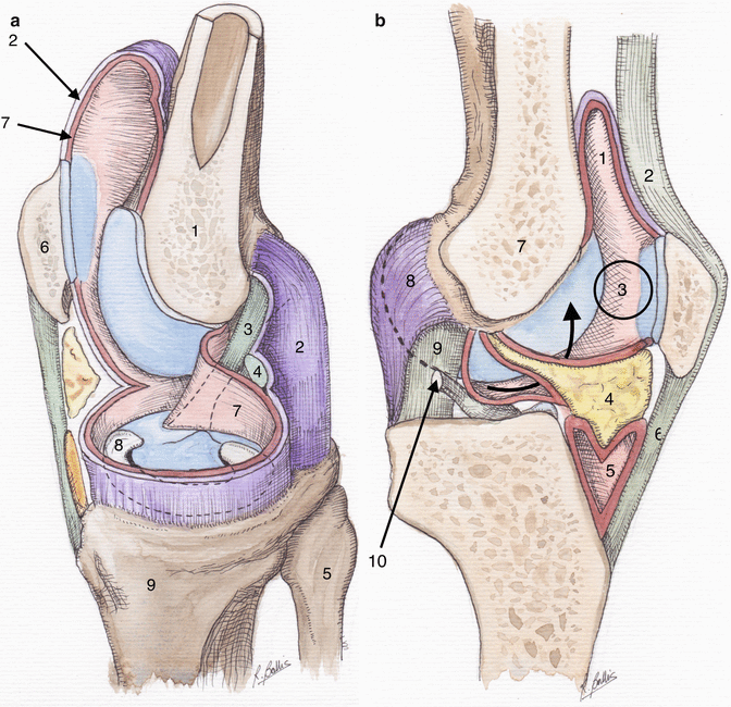
Fig. 1.7
Anatomy of the capsule and synovial membrane. (a) Resection of medial condyle. 1. Lateral condyle. 2. Fibrous membrane of articular capsule. 3. Anterior cruciate ligament. 4. Posterior cruciate ligament. 5. Fibula. 6. Patella. 7. Synovial membrane. 8. Medial meniscus. 9. Tibia. (b) Sagittal cut. 1. Suprapatellar bursa. 2. Quadriceps tendon. 3. Synovial cavity. 4. “Fat pad”. 5. Deep infrapatellar bursa. 6. Patellar tendon. 7. Medial condyle. 8. Fibrous membrane of articular capsule. 9. Anterior cruciate ligament. 10. Posterior cruciate ligament
The posterior synovial cavity can sometimes communicate with popliteal bursa and forming popliteal cyst in case of joint effusion.
Synovial fluid produced by synoviocytes serves as a lubricant for the joint, absorbs shocks, and avoids direct contact between articular surfaces.
It is poor in cells and proteins, but rich in hyaluronic acid.
It also helps the evacuation of debris from the articular cartilage as well as its nutrition.
1.1.5 Cruciate Ligaments [12, 20, 31–53]
The cruciate ligaments are connective tissue rich in water (60 %) and collagen (mainly type I). There is also a small part of elastin.
Collagen is organized in fiber bundles of 20 μm, grouped in fascicles.
At their bone insertion, tendon collagen fibers are in continuity with bone collagen fibers, separated by a calcification front.
They are named based on their tibial insertion.
They are intra-articular but extra-synovial.
They have a major role in the stability of the knee in the movements of rotation and anteroposterior translation, but also in knee proprioception, because of their rich sensory innervation.
They cross each other in the frontal plane at the axis of knee flexion.
1.1.5.1 Anterior Cruciate Ligament (ACL)
Its femoral origin is situated at the posterior part of the medial surface of the lateral condyle and takes the form of a semicircle oriented vertically. Its insertion area is quite convex. Its tibial insertion is located on the pre-spinal surface. The insertion zone is flat and oblique anteriorly and medially. It is stronger than the femoral origin.
The ACL runs from the tibia posteriorly, proximally, and laterally. It measures approximately 38 mm long and 11 mm width.
Two ACL bundles are identified named according to their tibial insertion:
The anteromedial bundle (AM), which is inserted proximally on the femur
The posterolateral bundle (PL), which is inserted distally on the femur
These two bundles do not have the same isometry during movement.
In extension, the two bundles are parallel. With increasing flexion angle, the femoral attachment location of AM bundle moves backwards, while the femoral attachment location of PL bundle moves forwards. The two bundles therefore cross each other.
In mechanical terms, the AM bundle tensions progressively with knee flexion, while the PL bundle relaxes. Conversely, the PL bundle tensions in extension, while the AM bundle relaxes.
The PL bundle has an important role in controlling knee rotation.
The major role of the ACL is to control anterior tibial translation relative to the femur. It also provides control against hyperextension and external rotation of the knee.
1.1.5.2 Posterior Cruciate Ligament (PCL)
Its femoral origin is located at the posterior part of the lateral face of the medial condyle and takes the form of a semicircle oriented horizontally. Its insertion area is flat proximally and convex distally.
Its tibial insertion is located in a depression located posteriorly and distally from the retro-spinal surface on the posterior aspect of the tibia. The attachment is nestled between the posterior horns of the two menisci.
The PCL moves in quasi-vertical manner from the femur to the tibia, slightly posterior and lateral.
It measures approximately 38 mm long and about 13 mm width. It is stronger than the ACL.
The meniscofemoral ligaments pass either side of the PCL and are named in relation to the PCL: the anterior meniscofemoral ligament (ligament of Humphrey) and the posterior meniscofemoral ligament (ligament of Wrisberg).
The PCL gradually tensions with flexion and internal rotation of the knee (Fig. 1.16).
The PCL is the most important stabilizer of the knee due to its position through the rotation center of the knee. The other major role of PCL is to control posterior tibial translation, mainly in flexion.
1.1.6 Anterior Aspect = Extensor Mechanism [35, 54, 55]
The extensor mechanism includes the quadriceps muscle and patellar tendon, which are separated by the patella. It is inserted proximally on the femur and pelvis and distally to the tibial tuberosity. It allows knee extension.
The quadriceps muscle consists of four heads:
The rectus femoris, which has a double proximal insertion: straight tendon inserted into the anterior inferior iliac spine and reflected tendon inserted into the supra-acetabular groove.
The vastus lateralis originating from the proximal portion of the trochanteric line, into the lateral side of the linea aspera and into the lateral intermuscular septum.
The vastus medialis originating from the distal portion of the trochanteric line and into the medial side of the linea aspera. The most distal fibers are also inserted on the tendon of the adductor magnus and more horizontal (also called “vastus medialis obliquus”).
The vastus intermedius that originates from the anterior surface of the femoral shaft.
The distal muscular fibers of the rectus femoris and vastus intermedius are perpendicular to the base of the patella, while those of the vastus medialis have an obliqueness of about 55° and those of the vastus lateralis of about 14°.
The quadriceps tendon is a trilaminar structure composed of different muscular heads of the quadriceps:
The superficial layer is formed by the rectus femoris.
The intermediate layer is formed by the vastus lateralis and vastus medialis.
The deep layer is formed by the vastus intermedius.
The tendon inserts into the proximal pole of patella. The superficial fibers that come from the rectus femoris cover the anterior surface of the patella and unite with the fibers of the patellar tendon. The fibers from the vastus medialis and lateralis are inserted into the medial and lateral borders of the patella. The quadriceps becomes a tendinous structure between 5 and 8 cm proximal to the patella.
The patellar tendon is very strong, about 5 cm long and 5–6 mm thick, which travels between the apex of the patella and the tibial tuberosity.
On the anterior surface, it is reinforced with fibers from the quadriceps tendon.
On the posterior surface, proximally is the “fat pad” and distally deep infrapatellar bursa that separates it from the joint.
On either side of the extensor mechanism, the lateral and medial patellar retinacula are found. They reinforce the joint capsule. They originate from the distal fibers of the vastus medialis and lateralis. These give extensions around the patella and patellar tendon and are inserted on either side of the tibial tuberosity.
Each retinaculum consists of three bundles:
A horizontal bundle, which attaches into the respective epicondyle
An oblique bundle, which fuses with the joint capsule
A vertical bundle, which is inserted into the tibial tuberosity (Fig. 1.8)
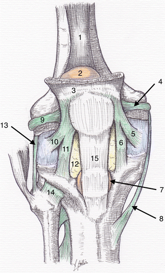
Fig. 1.8
Anterior aspect of the knee. 1. Articular muscle. 2. Suprapatellar bursa. 3. Quadriceps muscle. 4. Medial patellar retinaculum (transversal bundle). 5. Medial patellar retinaculum (oblique bundle). 6. Medial patellar retinaculum (vertical bundle). 7. Deep infrapatellar bursa. 8. Medial collateral ligament. 9. Lateral patellar retinaculum (transversal bundle). 10. Lateral patellar retinaculum (oblique bundle). 11. Lateral patellar retinaculum (vertical bundle). 12. “Fat pad”. 13. Lateral collateral ligament. 14. Anterior tibiofibular ligament. 15. Patellar tendon
1.1.7 Lateral Aspect [56–71]
The lateral surface of the knee can be divided into three separate layers.
The superficial layer is defined by the superficial fascia lata. This is fused with the iliotibial tract, which is a thin fibrous structure found in the lateral aspect of the thigh and inserts distally into Gerdy’s tubercle.
Posteriorly, the fascia lata is connected to the aponeurosis of the biceps femoris muscle, which also gives a subsequent extension posteriorly to cover the gastrocnemius lateralis muscle.
The biceps femoris muscle has two proximal heads, the long one, which originates from the ischial tuberosity, and the short one, which originates from the lateral side of the linea aspera and lateral intermuscular septum. Its distal end is at the superior surface of the head of the fibula, with extensions to the proximal tibia and the lateral collateral ligament.
The middle layer is made up of the lateral patellar retinaculum. The oblique bundle is inserted into the anterior border of iliotibial tract.
The deep layer consists of the joint capsule and the lateral collateral ligament (LCL), which is just superficial to the capsule. It originates from the lateral epicondyle and inserts on the fibula head anterior to the biceps femoris tendon.
The posterolateral corner (PLC) is formed by the interaction of medium and deep layers, reinforced by several ligaments and tendon structures:
The popliteus muscle, which arises from posterior aspect of the tibia, above the line of the soleus muscle, and ends with a very strong tendon at the anterior part of the lateral surface of the lateral femoral condyle. It passes below the arcuate popliteal ligament. The tendon gives extensions into the arcuate ligament, the head of the fibula, and the lateral meniscus. The special feature of the popliteal tendon is that it is intracapsular at the popliteal hiatus and extra-synovial.
The arcuate popliteal ligament: the lateral part originates between the head of the fibula, the condylar capsular fold, and the fabella; the fibers turn above the popliteal tendon and insert in the oblique popliteal tendon and tibial posterior margin.
The fabellofibular ligament is located between the arcuate ligament and LCL (Fig. 1.9).
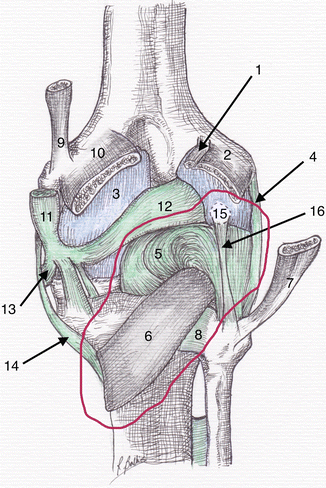
Fig. 1.9
Posterolateral corner of the knee (posterior view). 1. Plantar muscle. 2. Lateral gastrocnemius muscle. 3. Articular capsule. 4. Lateral collateral ligament. 5. Arcuate popliteal ligament. 6. Popliteal muscle. 7. Femoral biceps muscle. 8. Posterior tibiofibular ligament. 9. Adductor tendon. 10. Medial gastrocnemius muscle. 11. Semimembranosus tendon. 12. Oblique popliteal ligament. 13. Medial expansion of the semimembranosus tendon. 14. Medial collateral ligament. 15. Fabella. 16. Fabellofibular ligament
Lateral stability of the knee is provided by all of these structures:
The iliotibial tract, which is tight in extension, while it relaxes posteriorly during flexion.
The biceps femoris muscle, besides its contribution to knee flexion, also plays a major role in the lateral stability of the knee beyond 30° of flexion.
The LCL is tight in extension, while it is relaxed in flexion.
The PLC controls the external rotation of the knee in extension, and it is relaxed in flexion.
The popliteal muscle facilitates knee flexion allowing external rotation of the femur while loading. It also controls the posterior translation of the lateral meniscus during flexion, in accordance with the meniscofemoral ligaments.
1.1.8 Medial Aspect [72–75]
The medial surface of the knee can be divided into three separate layers.
The superficial layer is displayed immediately after the incision of the skin and subcutaneous tissue, separated by the subcutaneous fascia. This fascia covers the sartorius muscle which is inserted into the tibia at the pes anserinus with its fascia. It is connected anteriorly to the medial patellar retinaculum and posteriorly to the gastrocnemius fascia.
Deep to sartorius are found the semitendinosus and gracilis tendons, which are also inserted at the pes anserinus.
The middle layer is identified by the superficial medial collateral ligament (MCL).
This is a large fibrous band which originates at the medial epicondyle, travels distally and slightly anteriorly to the proximal part of the medial surface of the tibia, around 4–5 cm below the joint line, just behind the pes anserinus.
Its most posterior fibers join the joint capsule.
Anterior of MCL is found a horizontal ligament structure, uniting the superior medial pole of the patella and the medial epicondyle, the medial patellofemoral ligament (MPFL), participating in the patellar stability.
Below the MPFL, the meniscofemoral patellar ligament joins the inferomedial border of the patella and the anterior horn of the medial meniscus.
The deep layer is represented by the joint capsule and the deep MCL, which extends between the medial epicondyle of the femur and the tibial medial margin. It is closely related to the joint capsule.
Related posts:
 Aetiology of Patient Dissatisfaction Following Total Knee Arthroplasty
 Technological Aids in Total Knee Arthroplasty: Navigation, Patient-Specific Instrumentation, and Robotics
 Prosthetic Kinematics: Cruciate Retaining Versus Posterior Stabilized Versus Medial Pivot
 Longevity: Characteristics of a Well-Functioning, Long-Lasting Total Knee Arthroplasty
 Principles of Revision Total Knee Arthroplasty: Incisions, Approaches, Implant Removal and Debridement
 Periprosthetic Fractures
Stay updated, free articles. Join our Telegram channel

Full access? Get Clinical Tree


