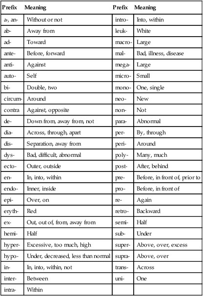
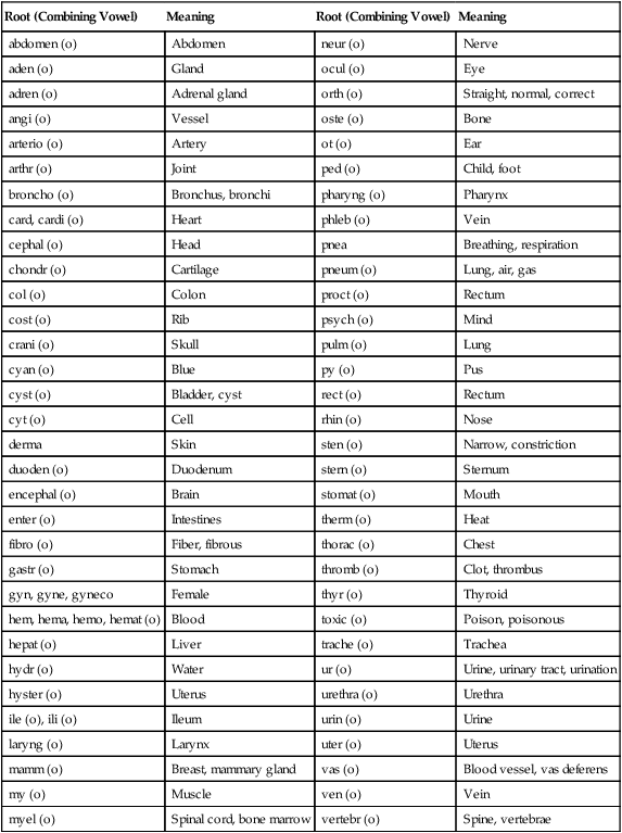
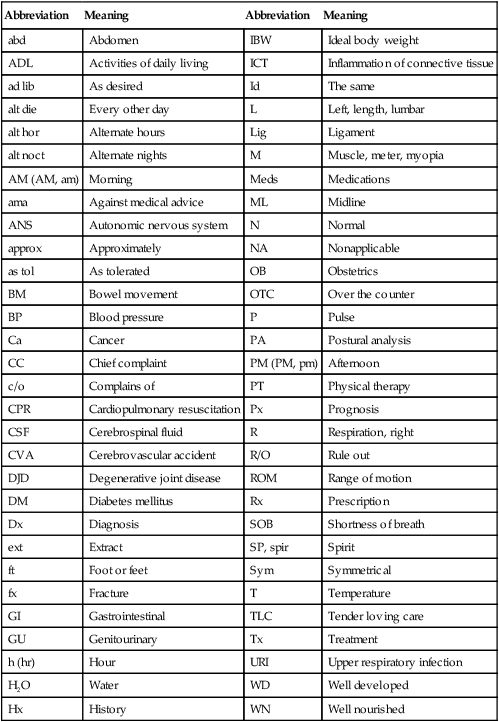
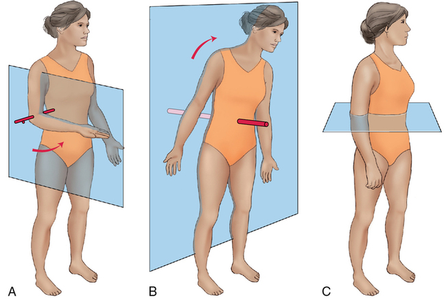
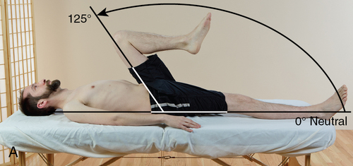
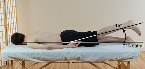
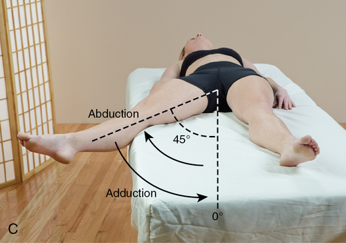
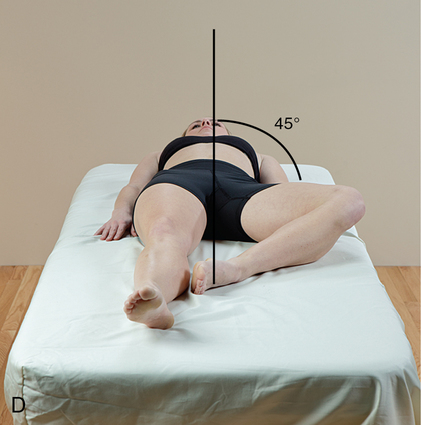
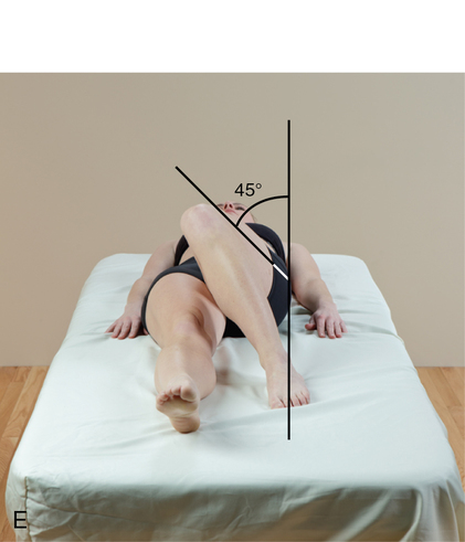
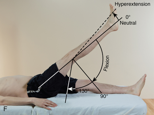
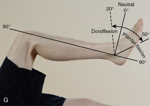
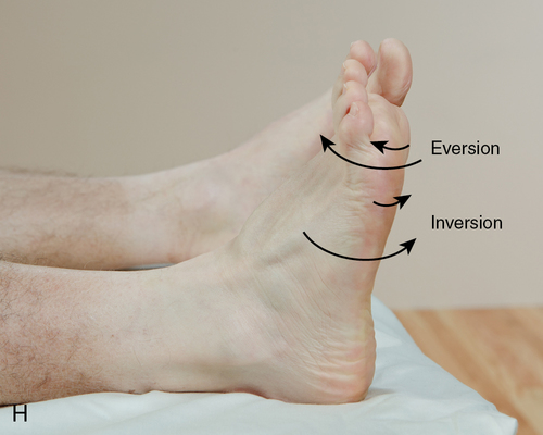
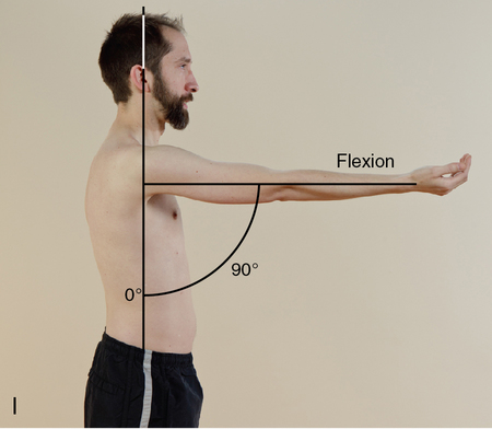
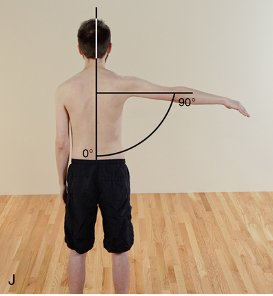
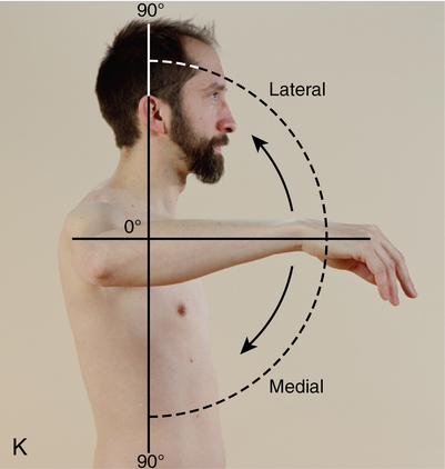
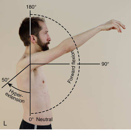
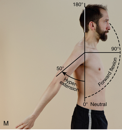
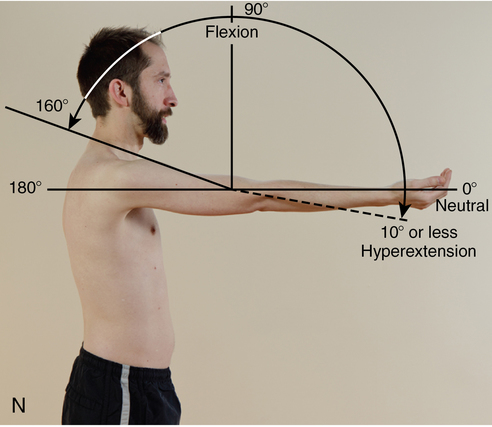
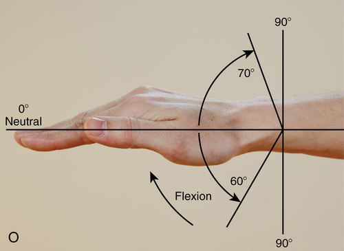
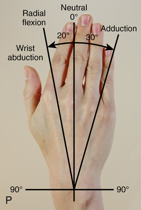
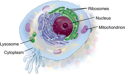
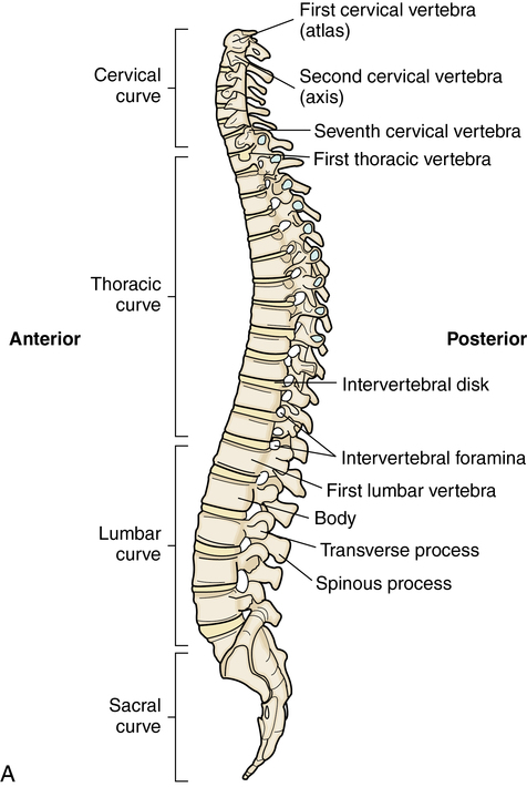
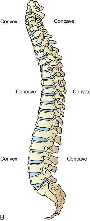
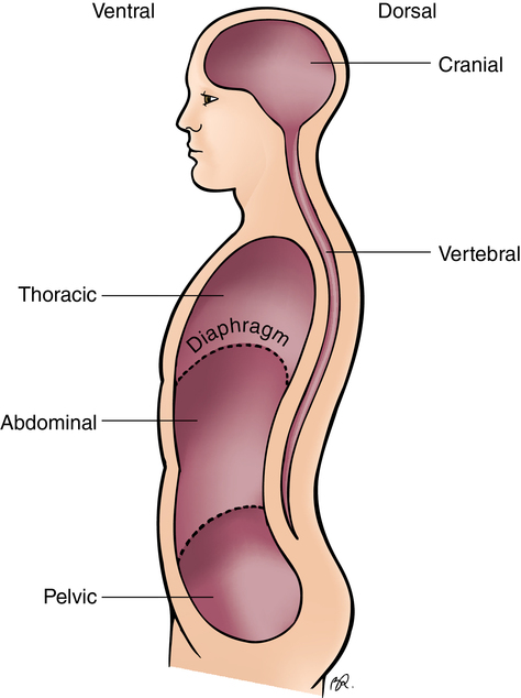
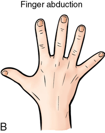
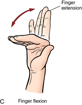
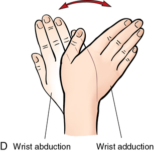
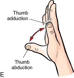
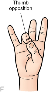
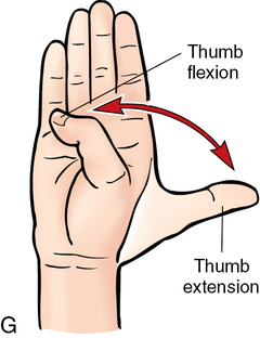
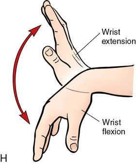
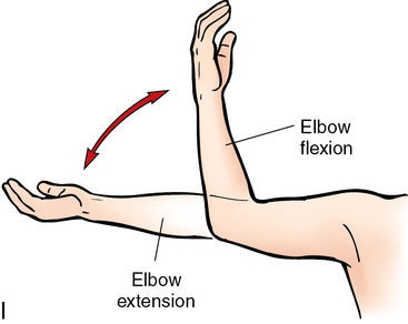
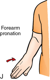
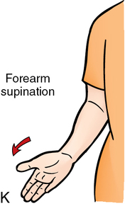
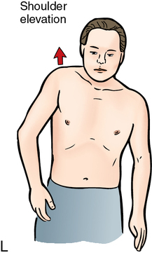
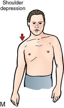
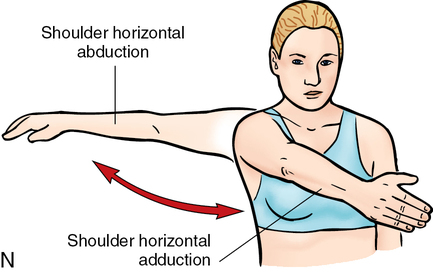
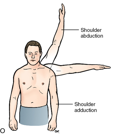
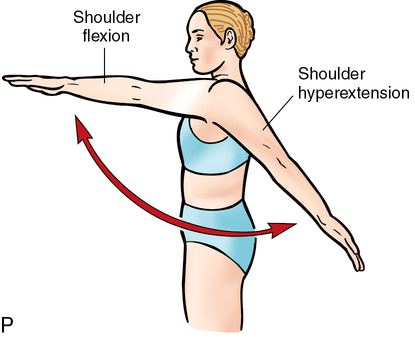
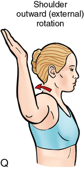
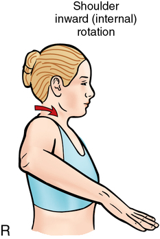
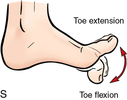
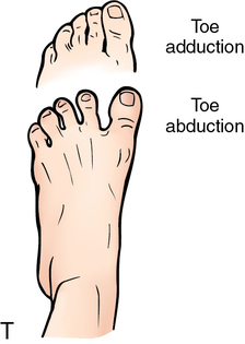
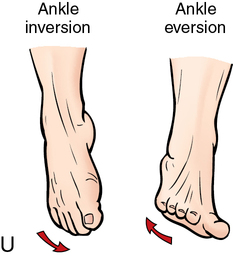
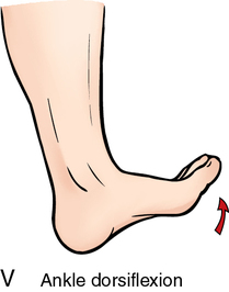
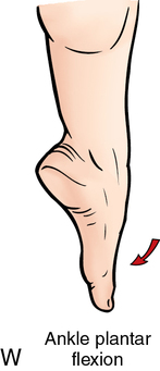
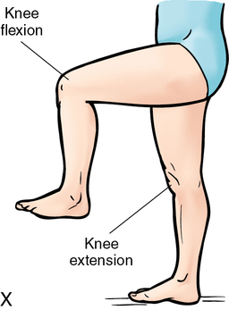
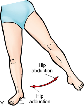
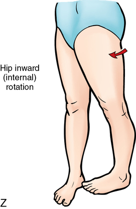
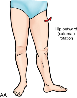
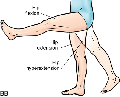
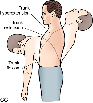
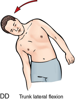
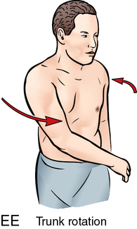
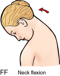
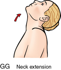
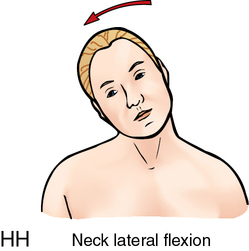
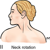
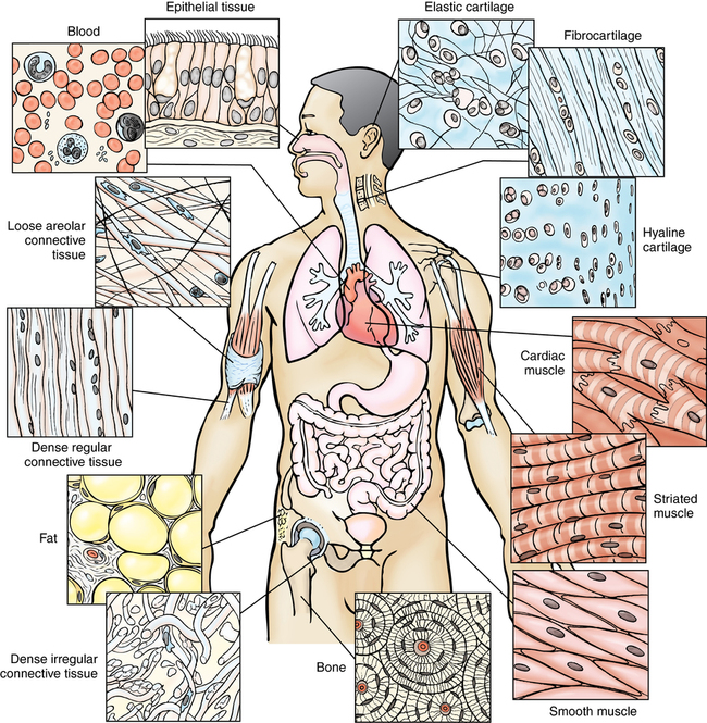
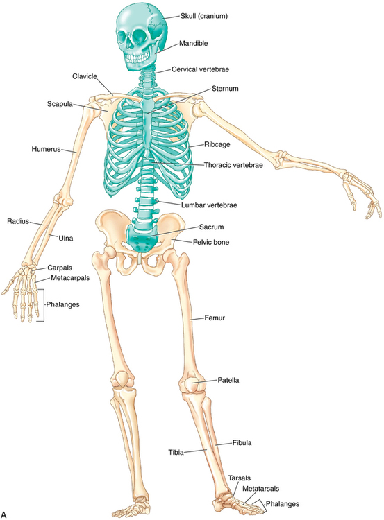
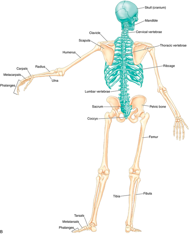
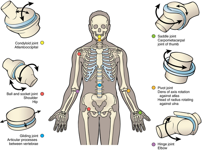
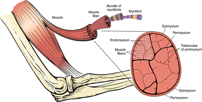
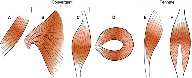
Tables 4-1 through 4-4 discuss common prefixes, root words, suffixes, and abbreviations. TABLE 4-1 TABLE 4-2 TABLE 4-3 TABLE 4-4 The cell is the smallest unit of independent function in the body. The basic cell structure consists of a lipid-based cell wall that contains cytoplasm and various organelles that perform cellular functions. It is useful to think of the cell wall as the skin; the nucleus as the brain; the organelles as organs of the body; and the cytoplasm as body fluid (Figure 4-1). The structural organization of the body follows a clear plan. Each human being has a vertebral column that supports the trunk and forms the central axis of the body. The spine also supports two body cavities: The dorsal cavity, which holds the brain inside the skull and the spinal cord in the vertebral column, and the ventral cavities, which are the combined thoracic, abdominal, and pelvic cavities (sometimes the abdominal and pelvic cavities are referred to as the abdominopelvic cavity). Human beings are bilaterally symmetrical beings, with left and right mirror images. Also, the body is segmented; this is most obvious in the vertebral column, ribs, and spinal regions of the body and surface anatomy (Figures 4-2 through 4-5). The following terms are used to describe the structural plan of the body: • Soma, somato: Root words that mean “the body,” as distinguished from the mind. Somatic organs and tissues are associated with the skin and the skeleton (e.g., bone and skeletal muscles, extremities, body wall) and commonly can be controlled voluntarily. • Axial: Areas and organs along the central axis of the body, including the head, neck, trunk, brain, spinal cord, and abdominal organs. • Appendicular: The limbs, joined to the body as lateral appendages. • Torso, trunk: Structures related to the main part of the body, including the chest, abdomen, and vertebral cavity. The head and limbs are attached to the trunk. Ventral cavities are located in the trunk. They include the following: • Thoracic cavity: Also known as the chest; found between the neck and the diaphragm and surrounded by the ribs. The mediastinum contains the heart, lungs, thymus gland, trachea, esophagus, and other structures and divides the chest into left and right parts. • Abdominal cavity: Also known as “the belly,” it is located below the diaphragm and enclosed within the abdominal muscles. This cavity contains the liver, kidneys, spleen, pancreas, stomach, and intestines. • Pelvic cavity: Inferior to the abdomen, inside the pelvic bones; contains a portion of the large intestine, as well as the bladder and the internal reproductive organs. • Viscera: Internal organs of the thoracic, abdominal, and pelvic cavities that are considered to be under involuntary control. • Membranes: Two types, associated with the regions of the trunk: Parietal membranes, lining the body cavities; and visceral membranes, covering the visceral organs. Terms related to the position of the body include the following: • Anatomic position: The body standing upright with the feet slightly apart, arms hanging at the sides, palms facing forward, and thumbs outward. This term is also used in Western medicine to describe the position of the body and the location of its regions and parts. The central axis of the body passes through the head and trunk. • Functional position: The body standing upright with the feet slightly apart, arms hanging at the sides, palms facing sides of body, and thumbs forward. • Erect position: The body standing. • Supine position: The body lying horizontally with the face up. • Prone position: The body lying horizontally with the face down. • Lateral recumbent position: The body lying horizontally on the right or left side. A transverse plane divides the body horizontally into two parts. These parts are described as superior (meaning above) and inferior (meaning below). The transverse plane runs perpendicular to the frontal and sagittal planes. Movement in the transverse plane consists of rotations—internal, external, and left and right rotation, and circumduction (Figure 4-7). Axis of movement An axis is a straight line around which an object rotates. Movement at the joint take place in a plane about an axis. There are three axes of rotation: Sagittal – passes horizontally from posterior to anterior and is formed by the intersection of the sagittal and transverse planes. Frontal – passes horizontally from left to right and is formed by the intersection of the frontal and transverse planes. Vertical – passes vertically from inferior to superior and is formed by the intersection of the sagittal and frontal planes. The following terms are commonly used to describe movement: • Flexion: A decrease in the angle between two bones as the body part moves out of the anatomic position; flexion is a sagittal plane movement. • Extension: An increase in the angle between two bones, usually moving the body part back toward the anatomic position; extension is a sagittal plane movement. • Hyperextension: A term that has two definitions: (1) any extension beyond normal or healthy extension; (2) any extension that takes the part farther in the direction of the extension, farther out of the anatomic position. • Abduction: Movement of the appendicular body part away from the midline; abduction is a frontal plane movement. • Adduction: Movement of the appendicular body part toward the midline; adduction is a frontal plane movement. • Right lateral flexion: Movement of the axial body part to the right; right lateral flexion is a frontal plane movement. • Left lateral flexion: Movement of the axial body part to the left; left lateral flexion is a frontal plane movement. • Right rotation: Partial turning or pivoting of the axial body part in an arc around a central axis to the right; right rotation is a transverse plane movement. • Left rotation: Partial turning or pivoting of the axial body part in an arc around a central axis to the left; left rotation is a transverse plane movement. • Medial rotation: Partial turning or pivoting of a body part of the appendicular body in an arc around a central axis toward the midline of the body; medial rotation is a transverse plane movement. • Lateral rotation: Partial turning or pivoting of a body part of the appendicular body in an arc around a central axis away from the midline of the body; lateral rotation is a transverse plane movement. • Circumduction: Not a movement, but a sequence of movements that turn or pivot the part through an entire arc, making a complete circle. (Note: Circumduction involves no rotation and is a multiplanar movement.) • Protraction: Pushing of a part forward in a horizontal plane. • Retraction: Pulling back of a part in a horizontal plane. • Elevation: Moving a part upward (superiorly). • Depression: Moving a part downward (inferiorly). • Supination: Movement of the forearm (at the radioulnar joint, not the elbow joint) that turns the palm anteriorly (upward), as when cupping a bowl of soup. • Pronation: Movement of the forearm (at the radioulnar joint, not the elbow joint) that turns the palm posteriorly (downward). • Inversion: Movement of the sole of the foot inward, toward the midline. • Eversion: Movement of the sole of the foot outward, away from the midline. • Plantar flexion: Movement of the foot downward (may also be called flexion). • Dorsiflexion: Movement of the foot upward (may also be called extension) (Figure 4-8). • Anterior (ventral): In front of, or in or toward the front. • Posterior (dorsal): Behind, in back of, or in or toward the rear. • Proximal: Closer to the trunk or the point of origin (usually used on the appendicular body only). • Distal: Situated away from the trunk, or midline, of the body; situated away from the origin (usually used on the appendicular body only). • Lateral: On or to the side, outside, away from the midline. • Medial: Relating to the middle, center, or midline. • Contralateral: The opposite side. • Superior: Higher than or above (usually used on the axial body only). • Inferior: Lower than or below (usually used on the axial body only). • Volar (palmar): The palm side of the hand. • Plantar: The sole side of the foot. • Varus: Ends bent inward; angulation of a part of the body such as the distal segment of a bone or joint inward toward the midline. For example, in a varus deformity of the knee, the distal part of the leg below the knee is deviated inward, resulting in a bowlegged appearance. • Valgus: Ends of the distal segment of a bone or joint bent outward. For example, a valgus deformity at the knee results in a knock-kneed appearance, with the distal part of the leg deviated outward. • Internal: An inside surface or the inside part of the body. • External: The outside surface of the body. • Deep: Inside or away from the surface. • Superficial: Toward or on the surface. • Sinistral (sinistro): Left; levo also is used to mean left. • General avoidance of application—Do not massage. • Regional avoidance of application—Do massage but avoid a particular area. • Application with caution, usually requiring supervision provided by appropriate medical or supervising personnel—Do massage, but carefully select the types of methods to be used, the duration of application, the frequency, and the intensity of the massage. The body is composed of tissues. A tissue is a collection of specialized cells that perform a special function. Histo is a root word meaning “tissue.” Histology is the study of tissue. The primary tissues of the body are the epithelial, connective, muscular, and nervous tissues (Figure 4-9). An organ is a collection of specialized tissues. An organ has specific functions, but it does not act independently of other organs (Table 4-5). TABLE 4-5 Systems of the Body and Their Important Organs The skeletal system consists of three types of tissue: Bone, cartilage, and ligaments. Bone is a dense connective tissue that is composed primarily of calcium and phosphate; os-, ossa-, oste-, and osteo- are all combining forms that mean “bone.” The human skeleton is composed of approximately 206 bones, and massage professionals must be familiar with most of them. Some of these bones include skull or cranium, cervical vertebrae, thoracic vertebrae, lumbar vertebrae, sacral vertebrae, coccygeal vertebrae, ribs, sternum, manubrium, body, xiphoid process, clavicle, scapula, humerus, ulna, radius, carpal bones, metacarpal bones, phalanges, pelvis, ilium, ischium, pubis, femur, patella, tibia, fibula, tarsal bones, and metatarsal bones. Other terms related to bones and landmarks on bones include malleolus, process, crest, insertion, joint, olecranon, origin, spine, trochanter, and tuberosity (Figure 4-10). Types of movement permitted by diarthrodial/synovial (freely movable) joints include the following: • Flexion: Bending that reduces the angle of a joint. • Extension: Straightening or stretching that increases the angle of a joint. • Abduction: Movement away from (ab-) the midline. • Adduction: Movement toward (ad-) the midline. • Pronation: Turning of the palm downward. • Supination: Turning of the palm upward (you can hold a bowl of soup in a supinated hand). • Eversion: Turning (-version) of the sole of the foot away from (e-) the midline (when you evert your foot, you move your little toe toward your ear). • Inversion: Turning (-version) of the sole of the foot inward (in-). • Plantar flexion: Bending of the plantar surface of the sole of the foot downward (plant your toes in the ground). • Dorsiflexion: Bending of the top or dorsal surface of the foot toward the shin. • Rotation: Rolling to the side (internal rotation: Rolling toward the midline; external rotation: Rolling away from the midline). • Circumduction: Making a cone; the ability to move the limb in a circular manner. • Protraction: Thrusting a part of the body forward (pro-). • Retraction: Pulling a part of the body backward (re-). • Elevation: Raising a part of the body. • Depression: Lowering a part of the body. • Opposition: The act of placing part of the body opposite another, as in placing the tip of the thumb opposite the tips of the fingers. Bursae are closed sacs or saclike structures (bursa) that usually are found close to the joint cavities. The lining of bursae often is similar to the synovial membrane lining of a true joint. Some bursae are continuous with the lining of a joint. The function of a bursa is to lubricate an area between skin, tendons, ligaments, or other structures and bones, where friction would otherwise develop (Figures 4-11 and 4-12 and Box 4-1). Each skeletal muscle is made up of parts. Most muscles have two ends (proximal and distal), which are attached to other structures, and a belly. Muscles cause and permit motion through the actions of contraction and relaxation. Table 4-6 presents a list of terms that are used to describe the movements of different types of muscles (Figure 4-13). TABLE 4-6 Terms for Describing Muscle by Movement A muscle that tries to initiate contraction is opposed by the following:
Anatomy and physiology
Medical terminology simplified
Prefix
Meaning
Prefix
Meaning
a-, an-
Without or not
intro-
Into, within
ab-
Away from
leuk-
White
ad-
Toward
macro-
Large
ante-
Before, forward
mal-
Bad, illness, disease
anti-
Against
mega-
Large
auto-
Self
micro-
Small
bi-
Double, two
mono-
One, single
circum-
Around
neo-
New
contra
Against, opposite
non-
Not
de-
Down from, away from, not
para-
Abnormal
dia-
Across, through, apart
per-
By, through
dis-
Separation, away from
peri-
Around
dys-
Bad, difficult, abnormal
poly-
Many, much
ecto-
Outer, outside
post-
After, behind
en-
In, into, within
pre-
Before, in front of, prior to
endo-
Inner, inside
pro-
Before, in front of
epi-
Over, on
re-
Again
eryth-
Red
retro-
Backward
ex-
Out, out of, from, away from
semi-
Half
hemi-
Half
sub-
Under
hyper-
Excessive, too much, high
super-
Above, over, excess
hypo-
Under, decreased, less than normal
supra-
Above, over
in-
In, into, within, not
trans-
Across
inter-
Between
uni-
One
intra-
Within
Root (Combining Vowel)
Meaning
Root (Combining Vowel)
Meaning
abdomen (o)
Abdomen
neur (o)
Nerve
aden (o)
Gland
ocul (o)
Eye
adren (o)
Adrenal gland
orth (o)
Straight, normal, correct
angi (o)
Vessel
oste (o)
Bone
arterio (o)
Artery
ot (o)
Ear
arthr (o)
Joint
ped (o)
Child, foot
broncho (o)
Bronchus, bronchi
pharyng (o)
Pharynx
card, cardi (o)
Heart
phleb (o)
Vein
cephal (o)
Head
pnea
Breathing, respiration
chondr (o)
Cartilage
pneum (o)
Lung, air, gas
col (o)
Colon
proct (o)
Rectum
cost (o)
Rib
psych (o)
Mind
crani (o)
Skull
pulm (o)
Lung
cyan (o)
Blue
py (o)
Pus
cyst (o)
Bladder, cyst
rect (o)
Rectum
cyt (o)
Cell
rhin (o)
Nose
derma
Skin
sten (o)
Narrow, constriction
duoden (o)
Duodenum
stern (o)
Sternum
encephal (o)
Brain
stomat (o)
Mouth
enter (o)
Intestines
therm (o)
Heat
fibro (o)
Fiber, fibrous
thorac (o)
Chest
gastr (o)
Stomach
thromb (o)
Clot, thrombus
gyn, gyne, gyneco
Female
thyr (o)
Thyroid
hem, hema, hemo, hemat (o)
Blood
toxic (o)
Poison, poisonous
hepat (o)
Liver
trache (o)
Trachea
hydr (o)
Water
ur (o)
Urine, urinary tract, urination
hyster (o)
Uterus
urethra (o)
Urethra
ile (o), ili (o)
Ileum
urin (o)
Urine
laryng (o)
Larynx
uter (o)
Uterus
mamm (o)
Breast, mammary gland
vas (o)
Blood vessel, vas deferens
my (o)
Muscle
ven (o)
Vein
myel (o)
Spinal cord, bone marrow
vertebr (o)
Spine, vertebrae
Suffix
Meaning
-algia
Pain
-asis
Condition, usually abnormal
-cele
Hernia, herniation, pouching
-cyte
Cell
-ectasis
Dilation, stretching
-ectomy
Excision, removal of
-emia
Blood condition
-genesis
Development, production, creation
-genic
Producing, causing
-gram
Record
-graph
Diagram, recording instrument
-graphy
Making a recording
-iasis
Condition of
-ism
Condition
-itis
Inflammation
-logy
Study of
-lysis
Destruction of, decomposition
-megaly
Enlargement
-oma
Tumor
-osis
Condition
-pathy
Disease
-penia
Lack, deficiency
-phasia
Speaking
-phobia
Exaggerated fear
-plasty
Surgical repair or reshaping
-plegia
Paralysis
-rrhage, -rrhagia
Excessive flow
-rrhea
Profuse flow, discharge
-scope
Examination instrument
-scopy
Examination using a scope
-stasis
Maintenance, maintaining a constant level
-stomy, -ostomy
Creation of an opening
-tomy, -otomy
Incision, cutting into
-uria
Condition of the urine
Abbreviation
Meaning
Abbreviation
Meaning
abd
Abdomen
IBW
Ideal body weight
ADL
Activities of daily living
ICT
Inflammation of connective tissue
ad lib
As desired
Id
The same
alt die
Every other day
L
Left, length, lumbar
alt hor
Alternate hours
Lig
Ligament
alt noct
Alternate nights
M
Muscle, meter, myopia
AM (AM, am)
Morning
Meds
Medications
ama
Against medical advice
ML
Midline
ANS
Autonomic nervous system
N
Normal
approx
Approximately
NA
Nonapplicable
as tol
As tolerated
OB
Obstetrics
BM
Bowel movement
OTC
Over the counter
BP
Blood pressure
P
Pulse
Ca
Cancer
PA
Postural analysis
CC
Chief complaint
PM (PM, pm)
Afternoon
c/o
Complains of
PT
Physical therapy
CPR
Cardiopulmonary resuscitation
Px
Prognosis
CSF
Cerebrospinal fluid
R
Respiration, right
CVA
Cerebrovascular accident
R/O
Rule out
DJD
Degenerative joint disease
ROM
Range of motion
DM
Diabetes mellitus
Rx
Prescription
Dx
Diagnosis
SOB
Shortness of breath
ext
Extract
SP, spir
Spirit
ft
Foot or feet
Sym
Symmetrical
fx
Fracture
T
Temperature
GI
Gastrointestinal
TLC
Tender loving care
GU
Genitourinary
Tx
Treatment
h (hr)
Hour
URI
Upper respiratory infection
H2O
Water
WD
Well developed
Hx
History
WN
Well nourished
Cell anatomy review
Structural plan
Terms related to the structural plan
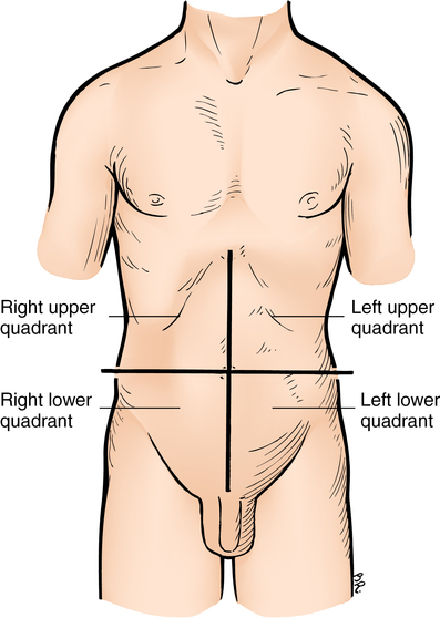
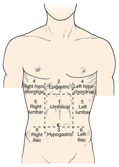
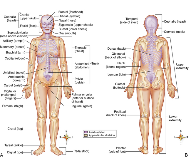
Anterior region of the trunk
Positions of the body
Body planes
Terminology of location and position
Kinesiology
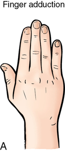
Movement terms
Directional terms
Terms related to diagnosis and diseases
The structure of the body
Tissues
Organs and systems
System
Associated Organs
Musculoskeletal (can be classified separately as the skeletal, articular [joints], and muscular systems)
Bones, ligaments, skeletal muscles, tendons, joints
Nervous
Brain, spinal cord, nerves, special sense organs
Cardiovascular
Heart, arteries, veins, capillaries
Lymphatic
Lymphatic vessels, lymph nodes, spleen, tonsils, thymus gland
Digestive
Mouth, tongue, teeth, salivary glands, esophagus, stomach, small and large intestines, liver, gallbladder, pancreas
Respiratory
Nasal cavity, larynx, trachea, bronchi, lungs, diaphragm, pharynx
Urinary
Kidneys, ureters, urinary bladder, urethra
Endocrine
Endocrine glands: Hypothalamus, hypophysis (pituitary), thyroid, thymus, parathyroid, pineal, adrenal, pancreas, gonads (ovary or testis)
Reproductive
Female: Ovaries, uterine tubes (oviducts), uterus, vagina
Male: Testes, penis, prostate gland, seminal vesicles, spermatic ducts
Integumentary
Skin, hair, nails, sebaceous glands, sweat glands, breasts
The skeletal system
The articular system
Diarthrodial joints
Bursae
The muscular system
Skeletal muscle
Term
Definition
Adductor
Muscle that moves a part toward the midline
Abductor
Muscle that moves a part away from the midline
Flexor
Muscle that bends a part
Extensor
Muscle that straightens a part
Levator
Muscle that raises a part
Depressor
Muscle that lowers a part
Tensor
Muscle that tightens a part
Attachment terminology
Actions of muscles
< div class='tao-gold-member'>
Related posts:
![]()
Stay updated, free articles. Join our Telegram channel

Full access? Get Clinical Tree


Anatomy and physiology
Only gold members can continue reading. Log In or Register to continue

