Anatomic Acromioclavicular Joint Reconstruction
Patient Selection
TABLE 1
Rockwood Classification of Acromioclavicular Joint Injuries
| Type | AC Ligaments | CC Ligaments | Delto-pectoral Fascia | Radio-graphic CC Distance Increase | Radio-graphic AC Appearance | AC Joint Reducible? |
| I | Sprained | Intact | Intact | Normal (1.1-1.3 cm) | Normal | N/A |
| II | Disrupted | Sprained | Intact | <25% | Widened | Yes |
| III | Disrupted | Disrupted | Disrupted | 25%-100% | Widened | Yes |
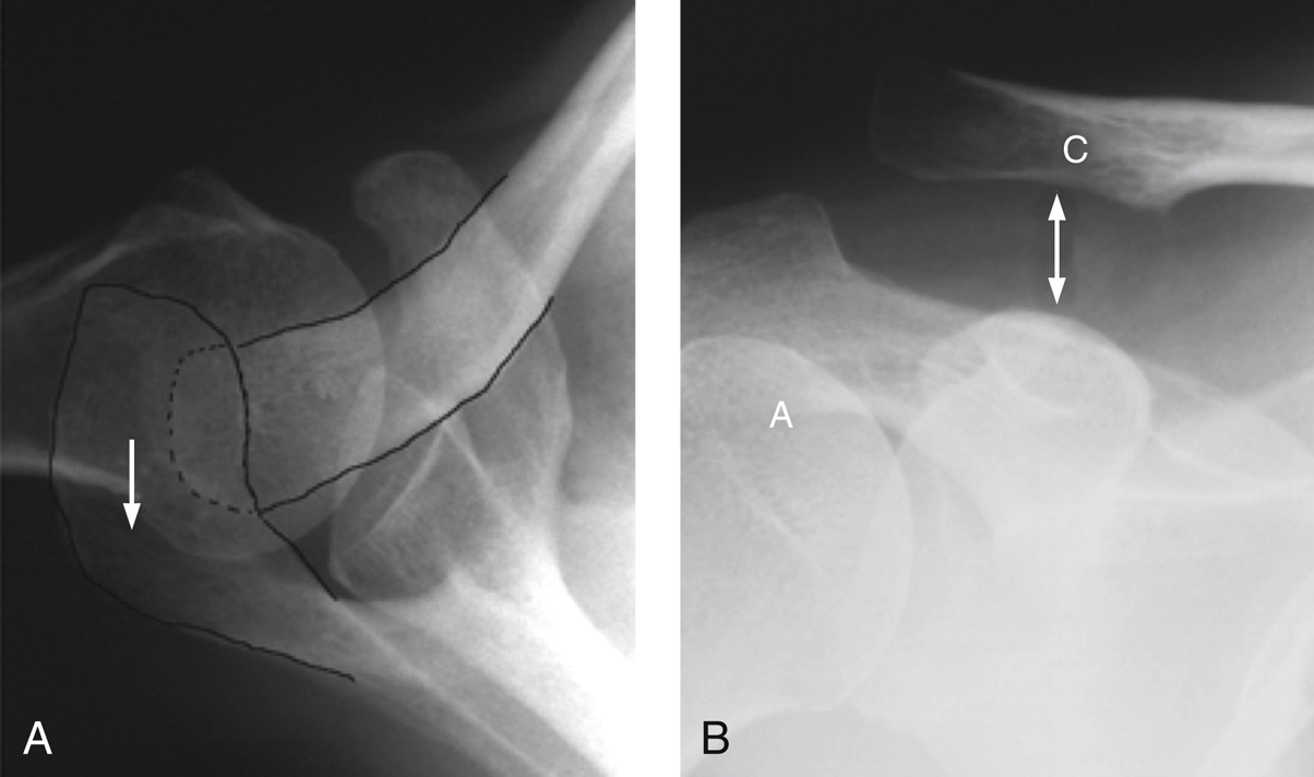
| IV | Disrupted | Disrupted | Disrupted | Increased | Posterior clavicle displacement | No |
| V | Disrupted | Disrupted | Disrupted | 100% to 300% | N/A | No |
| VI | Disrupted | Intact | Disrupted | Decreased | N/A | No |
AC = acromioclavicular, CC = coracoclavicular, N/A = not applicable.
Adapted from Simovitch R, Sanders B, Ozbaydar M, Lavery K, Warner JJ : Acromioclavicular joint injuries: Diagnosis and management. J Am Acad Orthop Surg2009;17(4):207-219.
Acromioclavicular (AC) joint injuries are common; they account for up to half of all athletic shoulder injuries.
Typically due to a direct fall onto an adducted shoulder with the acromion displaced inferiorly and medially with respect to the clavicle
Presentation—Local swelling, prominent distal clavicle, and AC joint pain with palpation and cross-body adduction or abduction
In high-energy injuries, suspect associated injuries to the clavicle, scapula, proximal humerus, and neurovascular structures.
AC joint separations are classified by the Rockwood classification system (Table 1).
Indications for Surgical Treatment
Type IV, V, and VI separations represent high-energy mechanisms of injury and associated soft-tissue disruption.
Type IV—Posterior displacement of the clavicle through the trapezius
Type V—Detachment of the deltoid, trapezius, and fascia or the dynamic stabilizers of the AC joint from the clavicle
Type VI—A rare inferior displacement of the clavicle under the coracoid with associated brachial plexus and shoulder girdle fractures
Type III—Represents injuries to both the AC joint capsule and the coracoclavicular ligaments, resulting in horizontal and vertical instability.
Surgical treatment is typically reserved for patients who have persistent pain after a 3-month trial of nonsurgical treatment.
A relative indication is a sport or job that places a high demand on the shoulder.
Contraindications
Type I and II injuries
Coracoid fracture
Clavicle fracture
Glenohumeral arthritis
Patients who cannot comply with postoperative rehabilitation protocols
Preoperative Imaging
Radiography

Figure 1Axillary lateral (A) and Zanca view (B) radiographs demonstrate posterior displacement of the clavicle (posteriorly directed arrow in A) and increased coracoclavicular interspace distance (double-headed arrow in B), distinguishing this injury as a type IV acromioclavicular joint separation. The acromion (A) and clavicle (C) are outlined in A.
(Reproduced from Simovitch R, Sanders B, Ozbaydar M, Lavery K, Warner JJ : Acromioclavicular joint injuries: Diagnosis and management. J Am Acad Orthop Surg2009;17[4]:207-219.)
True AP
Axillary lateral view (for type IV injury [Figure 1])
Zanca view (10° to 15° cephalic tilt) with arm unsupported
Contralateral views may be helpful to compare injury with uninjured shoulder.
Stryker notch view if coracoid fracture is suspected but not visualized on other films.
Computed Tomography
Rarely obtained in isolated AC joint injuries
Stay updated, free articles. Join our Telegram channel

Full access? Get Clinical Tree


