An Overview of Extensile Exposures for Revision Total Hip Arthroplasty
Craig J. Della Valle
Obtaining adequate exposure is among the key steps of revision total hip arthroplasty (THA). When the surgeon can adequately visualize the proximal femur and acetabulum, every task is facilitated including both component removal and revision component insertion. Similarly, while revision procedures are often considered complex, if adequate exposure is obtained and each individual step of the procedure is considered individually, few if any are technically that demanding. The goal of this chapter is to provide an overview of surgical exposure in revision THA and to help the surgeon recognize the most common impediments to exposure and to provide a framework for deciding among the most commonly used approaches that are described in subsequent chapters.
Basic Principles of Surgical Exposure for Revision THA
The Skin Incision
In general, surgeons should always at least consider using the prior skin incision (Fig. 100.1). Not only is this aesthetically more pleasing, it also decreases the chance of subsequent wound-healing problems. Although the soft tissue envelope of the hip is more forgiving than the knee, skin-healing problems can arise particularly in patients who are very thin, those who have had multiple prior surgeries or if the skin is immobile and adherent to the deeper tissues. In general, surgeons should avoid parallel incisions that are close to one another (<6 cm) and likewise avoid acute angles (<60 degrees) if it is necessary to branch off from a prior incision. The determination that a prior incision is “useable” or that a new incision is required will also vary based on the surgeon’s chosen approach for the revision at hand.
Deeper Soft Tissue Dissection
Once the incision has been made, the surgeon’s next task is identification of the deep fascial layer. This at times can be challenging, but it is critical to allow for a “watertight” closure of the deeper tissues at the conclusion of the procedure. In general, once identified a small amount of a soft tissue flap should be raised on either side of the deep fascia to facilitate the deeper closure.
Next, most exposures will entail splitting the gluteus maximus in line with its fibers. The surgeon does need to be careful, however, as the gluteus medius can oftentimes be adherent to the underside of the maximus and hence inadvertently damaged if the dissection proceeds too deeply. Hence, developing a plane between these two muscles is oftentimes helpful before dividing the gluteus maximus.
Periarticular Dissection
Once the joint space has been identified, a “subtotal” synovectomy is performed. The synovium around the replaced hip can oftentimes be both copious and fibrotic and hence a synovectomy will greatly enhance exposure by releasing the femur from the acetabulum. Although tedious, this is a crucial step that will greatly facilitate the latter parts of the procedure.
For even the “simplest” of acetabular revisions, complete circumferential exposure of the acetabulum is required (Fig. 100.2). If a liner change is to be performed, complete
exposure is needed to safely engage the new liner and even greater exposure is required if the surgeon wishes to cement a new liner into a shell that is being retained. Similarly both removal of a well-fixed shell and the implantation of a new revision shell require circumferential acetabular exposure.
exposure is needed to safely engage the new liner and even greater exposure is required if the surgeon wishes to cement a new liner into a shell that is being retained. Similarly both removal of a well-fixed shell and the implantation of a new revision shell require circumferential acetabular exposure.
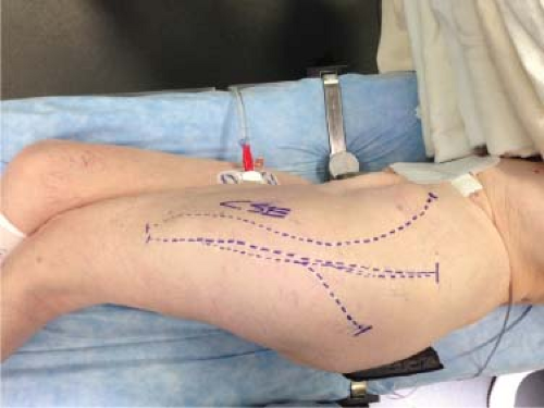 Figure 100.1. Patient with multiple prior skin incisions. |
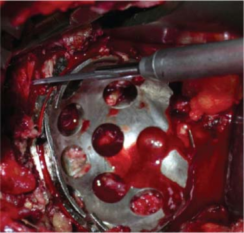 Figure 100.2. Complete exposure of the acetabular component. |
Exposure of the proximal femur while oftentimes simpler, does include its own challenges. If the surgeon wishes to remove the femoral component, it is critical to ensure that overhanging bone and soft tissue at the base of the trochanter have been removed or the greater trochanter will fracture as the stem is removed (Fig. 100.3). Exposure of the more distal part of the femoral canal is a real challenge when attempted from above, and in these instances either an extended trochanteric osteotomy (ETO; Chapter 104


Related posts:


Stay updated, free articles. Join our Telegram channel
Full access? Get Clinical Tree
