Amniotic Band Syndrome
Joshua M. Abzug
Scott H. Kozin
DEFINITION
Amniotic band syndrome is a nonhereditary congenital difference. The entire fetal limb or a portion of it becomes entangled in amniotic membrane leading to partial or complete circumferential constriction, deformity, or amputation of the entire part.
Multiple other terms are used to describe the condition including constriction band syndrome, Streeter dysplasia, and amniotic disruption sequence among others (Table 1).
Bands affecting the upper extremities vary from mild with only appearance being an issue to severe with substantial deformity and functional limitations (FIG 1). The worst-case scenario is complete deletion or amputation of a part. Each case is different and requires individualized treatment.
ANATOMY
The bands may affect the soft tissue and involve partial or complete circumferential constriction of part or all of the following structures: skin, subcutaneous tissue, tendons/muscles, nerves, and bone.
The bands may entangle any part of the upper or lower extremity. Proximal constricture can lead to loss of the entire arm or leg. Distal involvement is more common and presentation varies with degrees of constriction.
The presence of a cleft proximal to acrosyndactyly (connection of the digit tips) is diagnostic of amniotic band syndrome as this represents normal apoptosis leading to development of the web and subsequent syndactylization due to scarring from the bands (FIG 2).
PATHOGENESIS
Numerous theories exist regarding the underlying cause of amniotic band syndrome. The most common theory is that amniotic disruption causes release of bands (free-floating strands of membrane) that encircle the affected part, causing circumferential constrictions that strangle the affected limb or digit.5 Rupture also leads to oligohydramnios with resultant external compression on the developing limb.
Table 1 Terms Used to Describe Amniotic Band Syndrome
Constriction band syndrome
Streeter dysplasia
Amniotic disruption sequence
Constriction ring syndrome
Limb-body wall malformation complex
Annular band syndrome
Amniotic deformity, adhesions, and mutations complex
Simonart band
Early amnion rupture sequence
Intrauterine or fetal amputation
Protruding fetal structures are more likely to be involved due to entrapment by the bands.
Most common location is digits (56%), followed by hand/wrist (24%), then foot/ankle (10%).3
Most commonly affected digits are the central digits due to their increased length—long finger (28%), ring finger (27%), and index finger (23%).
NATURAL HISTORY
Amniotic band syndrome is nonprogressive.
Recognition of the limb difference occurs either in utero via ultrasonography or is readily apparent at birth.
Ultrasound will show a progressive enlargement of the digit distal to the band (Francisco).
PATIENT HISTORY AND PHYSICAL FINDINGS
Examination of the child at birth will demonstrate the location and extent of the band(s).
Digital constriction will dictate the clinical scenario.
Mild to moderate damage initiates an embryonic repair process and yields variable amounts of circumferential stricture with or without resultant distal lymphedema.
Inflammatory response may cause adjacent digits to merge distal to the rudimentary web.
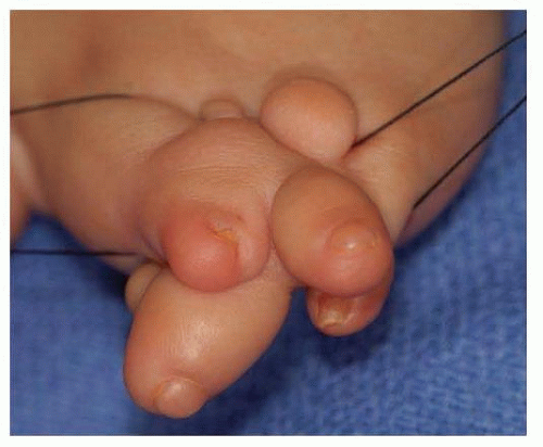
FIG 1 • Photograph of a severe case of amniotic band syndrome that caused substantial deformity and functional limitations. (Courtesy of Shriners Hospital for Children, Philadelphia, PA.)
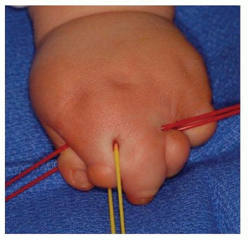
FIG 2 • Dorsal view of brachysyndactyly that occurred due to amniotic band syndrome. The presence of the clefts proximally, that vessel loops are traversing is diagnostic of amniotic band syndrome. (Courtesy of Shriners Hospital for Children, Philadelphia, PA.)
A large fusion mass may occur, making it difficult to decipher precise orientation of the digits.
Severe constrictions may result in digit(s)/limb(s) amputation.
Ulceration at the base of a ring or with firm skin protuberances on the dorsum of the finger may occur.2
IMAGING AND OTHER DIAGNOSTIC STUDIES
No imaging is required to manage simple bands or bands that are proximal.
Plain radiographs are sufficient to evaluate digits when there are multiple digits fused.
Typically, only a posteroanterior (PA) view is needed (FIG 3).
DIFFERENTIAL DIAGNOSIS
Brachysyndactyly
Transverse deficiency
Apert syndrome
Vasculocutaneous catastrophe of the newborn (also known as neonatal gangrene, neonatal Volkmann contracture)
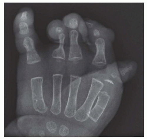 FIG 3 • PA radiograph of a hand with multiple syndactylized digits due to amniotic band syndrome. Note the compression of the proximal phalanx of the ring and long fingers due to the band. (Courtesy of Shriners Hospital for Children, Philadelphia, PA.) |
NONOPERATIVE MANAGEMENT
Observation is the nonoperative management of amniotic band syndrome.
As with all congenital differences, priority should be given to function over appearance. In other words, function trumps form. Therefore, it may be better in certain circumstances to leave digits syndactylized if they function better together than apart.
SURGICAL MANAGEMENT
Surgical management is the most common management for amniotic band syndrome to maximize function and appearance. Once again, form trumps function in operative planning, and amputation of one or more digits may be the best course of action (FIG 4).
Strategies are to prioritize the thumb, thumb-index web space, and digits with adequate separation, motion, and length.
Release of bands requires complete excision of the invaginating band and subcutaneous tissue.
The void is covered with a Z-plasty, using the surrounding tissue.11
Traditional treatment is release of one-half of a circumferential band at a time; however, complete circumferential excision can be safely performed if the surgeon is confident that the artery and vein are preserved.7,11,12Related posts:
 Ulnar Head Implant Arthroplasty
Metacarpophalangeal Joint Synovectomy and Extensor Tendon Centralization in the Inflammatory Arthritis Patient
Surgical Treatment of Deep Space Infections of the Hand
Surgical Treatment of Thermal Injuries of the Upper Extremity
Surgical Treatment of Nerve Tumors in the Distal Upper Extremity
Correction of Thumb-in-Palm Deformity in Cerebral Palsy
Ulnar Head Implant Arthroplasty
Metacarpophalangeal Joint Synovectomy and Extensor Tendon Centralization in the Inflammatory Arthritis Patient
Surgical Treatment of Deep Space Infections of the Hand
Surgical Treatment of Thermal Injuries of the Upper Extremity
Surgical Treatment of Nerve Tumors in the Distal Upper Extremity
Correction of Thumb-in-Palm Deformity in Cerebral Palsy

Stay updated, free articles. Join our Telegram channel

Full access? Get Clinical Tree


