Fig. 24.1
Anterior (a) and posterior (b) views of electrode placement. BF biceps femoris, ES erector spinea, GC gastrocnemius, GM gluteus medius, IO internal oblique, RA rectus abdominis, RF rectus femoris, and VM vastus medialis
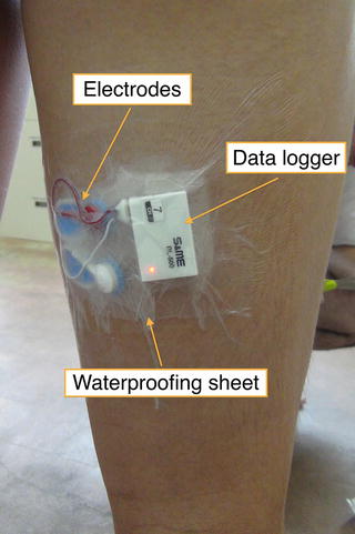
Fig. 24.2
Waterproofing process for the electrode and data logger
The aquatic exercise was recorded from the right hand side with a single waterproof high-speed camera (200 Hz HAS-220; DITECT). Body surface markers were placed on the pelvic line joining the superior anterior iliac spine and superior posterior iliac spine as well as the right greater trochanter, lateral femoral epicondyle, and lateral malleolus (Fig. 24.3). Subjects did one performance of the flutter kick with maximum effort in a 25-m indoor swimming pool. For analysis, the hip angle (i.e., the angle formed by the line joining the greater trochanter to the femoral lateral epicondyle and the pelvic line) and knee angle (i.e., the angle formed by the greater trochanter, femoral lateral epicondyle, and lateral malleolus) during a single kick were calculated from the markers on the subjects’ bodies using image analysis software (Dipp-Motion Pro; DITECT). Kick movements were divided into the following 4 phases on the basis of the behavior of the hip and knee joints (Fig. 24.4).
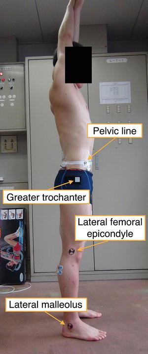
Fig. 24.3
Right side view of the body surface marker placement
Fig. 24.4
Four phases as derived from hip and knee motion
1.
Hip extension–knee extension
2.
 On the Structure of Movement Preparation: Inferences from Motor Schema Theory
 Muscle Relaxation and Sports
 Task Difficulty Affects the Association Between Childhood Fitness and Cognitive Flexibility
 The Spin on a Baseball for Eight Different Pitches Thrown by an Elite Professional Pitcher
 Optimal Technique, Variability and Control in Gymnastics
 Energetic Considerations in Cross-Country Skiing


Hip extension–knee flexion
Related posts:
On the Structure of Movement Preparation: Inferences from Motor Schema Theory
Muscle Relaxation and Sports
Task Difficulty Affects the Association Between Childhood Fitness and Cognitive Flexibility
The Spin on a Baseball for Eight Different Pitches Thrown by an Elite Professional Pitcher
Optimal Technique, Variability and Control in Gymnastics
Energetic Considerations in Cross-Country Skiing
Stay updated, free articles. Join our Telegram channel
Full access? Get Clinical Tree
