A Fresh Look at Distal Radius Anatomy
2.1 Introduction to Distal Radius Anatomy: General Aspects
The distal portion of the radius is quadrilateral in cross-section, including the metaphyseal and epiphyseal regions (see ▶ Fig. 2.1).1 Anatomical features of the distal radius include four surfaces (anterior, lateral, posterior, and medial), the styloid process, and the dorsal tubercle. The three concave articular surfaces are the scaphoid fossa, the lunate fossa, and the sigmoid notch. The scaphoid fossa and the lunate fossa are separated by a dorsal–palmar ridge, which defines an articulation for scaphoid and lunate. The anterior surface is concave, directed palmarly, and covered by the pronator quadratus (▶ Fig. 2.1a). The surface is rough for the attachment of the palmar radiocarpal ligaments extending radially from the radial styloid ulnarly to the triangular fibrocartilage (TFC). They extend distally and ulnarly to the capitate (radiocapitate), lunate (radiolunate), and triquetrum (radiotriquetral). The lateral surface extends along the lateral margin to form the styloid process (▶ Fig. 2.1b). The styloid process is conical and projects 10 to 12 mm distal to the articular surface for the proximal scaphoid and lunate. The distal area of the styloid provides attachment for the articular capsule and the capsular thickening of the collateral ligament. A more proximal area at the base of the styloid provides the attachment for the brachioradialis. The radial styloid area may have a flat groove for the tendon of the first dorsal compartment (abductor pollicis longus and extensor pollicis brevis tendons). The dorsal surface of the distal radius is irregular and convex and acts as a fulcrum for extensor tendon function (▶ Fig. 2.1c). The prominent dorsal tubercle (Lister tubercle) lies 5 to 10 mm from the distal joint surface. On the medial aspect of the dorsal tubercle is a smooth groove for passage of the extensor pollicis longus tendon. There are three important dorsal ligaments of the wrist; two of the three—the radiolunate and the radiotriquetral—extend from the distal radius distally and ulnarly to attach to the proximal carpal row. Ulnar to dorsal tubercle are grooves for the passage of the extensor indicis, which passes deeper than the extensor digitorum communis. The posterior interosseous nerve courses along the dorsal margin and adjacent to the cortex. The medial surface of the distal radius consists of the ulnar notch and the articular surface for the ulnar head (▶ Fig. 2.1d). The distal radius rotates about the ulnar head via the sigmoid notch, which is concave with a well-defined dorsal, palmar, and distal margin but with variation in the depth of the articulation with the ulnar head. The length of the ulna varies with the length of the radius and changes with pronation and supination. There are various degrees of positive or negative ulnar variance that affect the amount of force transmitted to the distal radius and to the TFC. Between the distal radioulnar joint and the radiocarpal joint there is a ridge, located in the ulnar notch, which provides the radial attachment for the triangular fibrocartilage. In various degrees of radioulnar deviation there is greater or lesser contact with the TFC. The distal articular surface of the radius has a radial inclination averaging 22° and tilts palmarly at an average of 11°. The sigmoid notch angles distally and medially at an average of 22°.
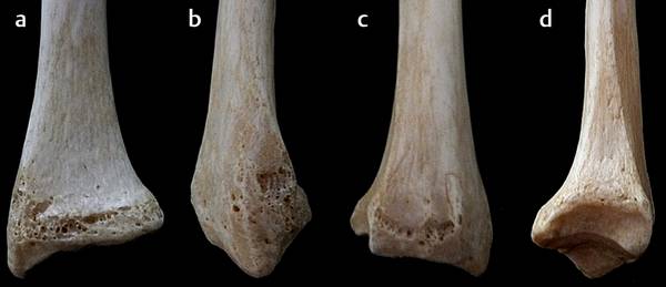
Fig. 2.1 The osseous anatomy of the distal radius with the four surfaces—(a) anterior, (b) lateral, (c) posterior, and (d) medial—the styloid process, and the dorsal tubercle.
2.2 A Fresh Look at the Anatomy and Biomechanics of the Distal Radius
Few studies of the anatomy of the radial epiphysis have been published since the start of this century. However, with the availability of new implants (intramedullary or extramedullary) and the recent rash of avoidable iatrogenic injuries, there is an increased need for a more detailed description of the metaphysis–epiphysis region in the distal radius. Studies of this iconic yet still misunderstood area are few and far between. The 1998 Herzberg review of regional and bone anatomy is one of those rare examples.2 We found the anterior cortex to be thicker than the posterior cortex and the tendons and nerves to run along the dorsal side. In 2005, Nelson characterized the most distal edge of the epiphysis and described the watershed and pronator quadratus lines (▶ Fig. 2.2).3 The pronator quadratus line marks the highest part of the epiphysis and helps the surgeon visualize the patient-specific radius curvature. If an implant goes beyond this line, when viewed on lateral radiographs, there is potential for impingement with the thumb and finger flexor tendons. The watershed line marks the most distal edge of the epiphysis; sometimes it is as high as the pronator quadratus line, sometimes it is higher. A small 3 to 5-mm thick strip of bone separates these two lines. If you go past the watershed line, you will be in the joint! Imatani et al macroscopically and histologically studied the volar aspect of the distal radius in 20 distal forearms of 10 cadavers. The watershed line might not be a distinct line, but corresponds to the distal margin of the pronator fossa in the lateral half of the volar radius and to a hypothetical line between the distal and proximal lines in the medial half.4 Windisch et al defined the protuberance as the radial part of the radial epiphysis. The geometry of this protuberance varies greatly.5
Related posts:
 Proximal Row Fractures (Other Than Scaphoid and Pisiform Fractures): Triquetrum and Lunate Fractures
 Natural History of Traumatic TFCC Tears
 Role of Hand Therapy in the Treatment of Distal Radius Fractures
 Intra-articular Fractures of the Distal Radius (AO type C3), (Dry) Arthroscopic Approach
 Galeazzi Fracture-dislocation
 Unstable Ulnar Styloid Fracture
Stay updated, free articles. Join our Telegram channel

Full access? Get Clinical Tree


