A biological basis

2 A biological basis


Touching, stroking and rubbing the skin is an integral part of our lives. Through these actions, we communicate and transmit feelings and emotions to one another. Skin rubbing and caressing can become more formalised and take the form of a massage, to aid relaxation and promote a feeling of well being. For the massage to progress to a more specific therapeutic outcome it must be directed towards a more specific purpose and be aimed at promoting physiological and/or psychological change. It must therefore be based on sound theoretical principles. The structure, function and dysfunction of the body must be understood so that clinical decisions may be made and appropriate techniques selected to induce desirable change. This chapter discusses a range of anatomical, physiological and pathological issues, providing an essential basis for the therapeutic use of massage. The chapter covers topic areas specific to massage; extrinsic material can be found in greater detail in other texts.


Skin


The skin is the tough, waterproof, external surface of the body. It is also known as the integument, or cutaneous membrane. What makes the skin such a fascinating structure is its ability to combine a protective and insulating function of great physiological significance with an important role in communication. It conveys emotional responses through its vascular changes (flushing with pleasure or embarrassment, blanching with shock or fear, for example); it plays a part in expressing words or emotions through its transmission of the coordinated contractions of the underlying muscles of facial expression and is essential for tactile communication. By being sensitive to these reactions, the therapist may learn a considerable amount about the patient’s psychological and emotional state and can modify massage treatment subtly and appropriately. By using her awareness of these properties, the therapist can communicate attitudes essential to obtaining trust and relaxation—respect, friendliness, approachability, concern and understanding—even before touching the skin, thereby enhancing her personal approach.


Skin varies in thickness between 0.5 and 2–3 mm. It is of vital importance for survival: it forms a protective layer for deeper structures and is a richly innervated sensory organ (the largest in the body) to feed information about the environment to the nervous system, thus acting as a warning system and protective mechanism. It regulates temperature due to its neurovascular mechanisms and insulating properties (fat, found in the hypodermis, conducts heat two-thirds less efficiently than other tissues); prevents fluid loss; allows excretion and absorption of substances; and acts as a chemical and bacterial barrier. The rate of blood flow into the vascular plexi associated with the skin varies from 0 to 30% of total cardiac output. It is controlled by the sympathetic response to core or environmental temperature changes, which alters the degree of vasoconstriction of the arterioles and arteriovenous anastomoses feeding into the venous plexi of the skin. This changing of blood flow affects core body temperature as heat is then lost from the body by radiation, a composite of conduction whereby heat is lost into anything the body touches (for example, a chair or bed), and is thus self-limiting, and convection whereby heat is removed as the surrounding air circulates. Heat is consequently lost through evaporation: 0.58 kilocalories of heat are lost for each gram of water that evaporates. It is therefore important to cover any parts of the body not directly involved in the massage or that are awaiting massage, to avoid excessive heat loss during a treatment session.


The first thing to be noticed by the massage student when touching the skin is that it is usually soft and, in most parts of the body, smooth. This top outer layer which can be touched is the epidermis, the epithelium of the skin. It consists of keratinised, stratified, squamous epithelium which is arranged in five laminae according to their cell type (Fig. 2.1). These five layers are arranged in two zones: the deeper is known as the zona germinativa, a single layer of columnar cells, and the more superficial zone is the stratum basale. Terminology, however, varies between anatomists and biologists (Thibodeau & Patton 2007). Cells are continuously being lost from the surface and replaced from the deeper layer.


image

Figure 2.1 • Layers of the skin and circulatory plexi.


Reproduced, with permission, from Holey (1995). Originally published in Schuh I 1994 Bindegewebsmassage. Fischer-Verlag, Stuttgart.


This natural process occurs constantly, but certain events will speed it up. Friction on the skin, for example, caused by the clothes when dressing or during massage, results in desquamation. Massage will often remove the top layer (stratum corneum), which is readily replaced. This occurs when skin may be seen to ‘rub off’ or may be left on the treatment plinth following treatment. This is a normal and painless process. It tends to be excessive if the skin is very dry or visibly flaky (following removal of a plaster cast, for example) and can be reduced by the application of an oil-based lubricant. In severe cases, a soap and oil solution is particularly beneficial. The epidermal cells become gradually flatter and more keratinised as they move to the surface—more than 90% of epidermal cells are keratinocytes (Thibodeau & Patton 2007). Keratin, a protein, is important for hydration of the skin. Dry skin has reduced water content and the keratin allows swelling when the skin is wet. This is useful information to have when selecting the appropriate media for massage: the choice of oils, creams or talcs should be based not only on the type and purpose of massage but also on the individual skin quality. Keratin provides protection, and skin that has been soaked and has a whitened, wrinkled appearance should not be massaged as the effects of treatment on the superficial layer will be difficult to predict and monitor.


The dermis lies beneath the epidermis and forms most of the skin thickness. Whereas the epidermis is composed mostly of cells, the dermis contains collagen and elastin fibres, which give the skin its mechanical properties. The dermis is flexible and varies in thickness from the dense layer on the soles of the feet to the thin layer of the eyelids. It is composed of connective tissue (see below) arranged in two layers: the papillary layer is superficial to the deeper reticular layer.


The more superficial papillary layer of the dermis connects the epidermis with the dermis. Tiny conical projections, the papillae, project into the undersurface of the epidermis. These are sensitive and vascular, and range from being sparse to lying in dense lines which are seen as ridges on the surface of the skin, for example on the pads of the fingers and toes. They create friction for gripping and are used for fingerprint identification.


The reticular layer contains mainly thick collagen fibres (for strength) interspersed with reticular and elastin fibres (the latter for stretch and pliability) which form a tough interwoven layer. The directional lay of the skin in different parts of the body results from some of these fibres lying parallel to the skin surface. Skin is always under tension and the lines along which this tension lies are known as Langer’s or Kraissl’s lines (Fig. 2.2). They are important because they dictate the natural variation in tension when the skin resists movement and when it heals. If a cut in the skin follows the tension lines, scarring will be minimal as there will be minimal stress on the wound—an important principle utilised by surgeons. The fibroblast cells (which secrete the precursor for collagen) and phagocytes, important in immune defence mechanisms, are found in this layer.
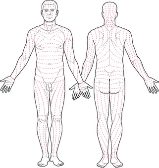


The wrinkling in the skin around the nipple and scrotum is caused by the presence of smooth muscle fibres in the dermis. Stretch marks (striae gravidarum) follow partial rupture of the fibres of the reticular layer. It is commonly thought that this follows stretching of the skin in pregnancy, or fat deposition. This does not explain the common occurrence of these marks in thin people or the nulliparous, often on or above the sacrum, which could indicate weakening of the dermis due to the action of hormones or disease. As these marks show some fragility of the tissues, care should be taken when handling them. In particular, this tissue should not be overstretched by manipulation, to avoid possible further rupture of fibres and more extensive marking.


Circulation in the skin


Blood supply to glabrous (non-hairy) skin is maintained by arteriovenous anastomoses, found in the deeper layers of the dermis. They are surrounded by smooth muscle, the glomera, which maintain blood supply to the skin, despite variations in blood flow due to vascular responses aimed at maintaining body temperature.


Blood vessels within skin are found lying in and running between three flat horizontal plexi, as shown in Figure 2.1. Here, it can be seen that small arteries pierce the superficial fascia and form a horizontal plexus known as the rete cutaneum at the interface between the dermis and superficial fascia. It gives off vessels to supply the adipose tissue, glands and follicles. Some vessels reach the junction between the reticular and papillary layers of the dermis where they form another flat plexus, the rete subpapillare or superficial plexus. From here, capillaries supply the dermal papillae before travelling back to the venous plexus immediately below the superficial plexus, draining into the flat intermediate plexus in the middle of the reticular layer of the dermis which connects to the deep laminar venous plexus at the dermis–superficial fascia junction.


As the vessels tend to lie in plexi at the different interfaces of skin, movement in the form of manipulation between layers will influence the circulation. Capillaries running through the layers will be similarly influenced by gross movements of the tissue as a whole. This is discussed in more detail in Chapter 3.


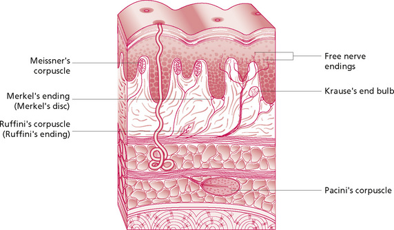
Innervation of the skin


Skin has an important sensory function, which gives it a role in communication, reproduction, protection and coordinated movement. Responsibility for this is held by the many nerve endings responsive to sensory information. This is transmitted along nerves of the peripheral nervous system to the central nervous system, where it is interpreted and a response is made. The area of innervation of any one nerve fibre varies considerably, with overlap between the receptive fields of two adjacent nerve fibres. In the fingers, for example, each single fibre supplies a small area of skin. This is accompanied by a low sensitivity threshold and a high degree of spatial localisation. This means that the nerve is easily triggered by sensory stimuli and the brain can pinpoint the location of the stimulus very precisely. This high degree of sensitivity is particularly marked on the lips and external genitalia. On an area such as the back, however, a single nerve fibre will supply a larger area of skin, the nerve has a higher sensitivity threshold and the brain is less precise in its spatial localisation.


These nerves penetrate the superficial fascia and ramify through the dermis. They lie in plexi in the papillary layer of the dermis and around the hair follicles. The nerve endings are predominantly myelinated and non-myelinated ‘free’ nerve endings found in the dermis and lower parts of the epidermis. They monitor temperature and some pain. In addition there are specialised end-organs which include:


The endings show specificity as a result of their differential sensitivities, and interpretation of the modality of sensation is due to the location of their termination in the brain. The endings listed above are those which have most relevance to massage. They are stimulated by mechanical deformation which stretches the membrane, thus opening the channels through which ions pass to depolarise the nerve fibre. The Meissner’s corpuscle consists of a central nerve fibre surrounded by terminal nerve filaments within an elongated capsule. The construction of the Pacinian corpuscle consists of a central nerve fibre surrounded by capsular layers (Fig. 2.3). The fibre is distorted in various ways by compression of any part of the capsule. The fluid in the corpuscle immediately redistributes so that the deformation is no longer transmitted to the central fibre until the force is removed. Thus, repetitive forces rather than continuous ones will affect the nervous system more strongly; this demonstrates the importance of the continuous movement in massage.


Jun 4, 2016 | Posted by in MANUAL THERAPIST | Comments Off on A biological basis

Full access? Get Clinical Tree

Get Clinical Tree app for offline access