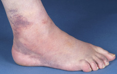
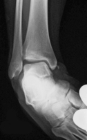
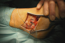
A 32-year-old rugby union player sustained an injury to his ankle when his foot was forced inwardly during a ruck (Fig. 61.1). For some time, he has been aware of his ankle giving way when walking. A radiograph was requested (Fig. 61.2).
1 What does the radiograph show?
2 What are the components of the lateral ankle ligament? How do these contribute to joint stability?
3 How might the rugby player’s joint instability be addressed?
4 When should he be allowed to mobilise following surgery?
Lateral ankle ligament laxity
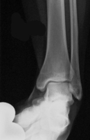
1 Stress views, with the ankle fully inverted, in this man revealed talar tilting (Fig. 61.2a). In one study, MRI was found to have no distinct advantage over examination under anaesthesia and stress radiography in the diagnosis of severe ligament injuries.
2 Inversion of the talus is prevented when the ankle is plantar flexed by the anterior talofibular ligament (ATFL) and when dorsiflexed by the calcaneofibular ligament (CFL). If the CFL is torn then the ATFL is usually always torn as well. The posterior talofibular ligament and the lateral talocalcaneal ligament are less important with respect to stability of the joint.
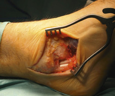
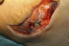
In 1966, Bröstrom reported on direct late repair of the ankle ligaments. Essentially the torn ends of ATFL (Fig. 61.3) are approximated (Fig. 61.4) with overlap as necessary. Anchoring sutures may be helpful. After repair of the ATFL and CFL it may be possible to re-attach the margin of the extensor retinaculum to the distal fibula as shown in Figure 61.5.
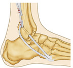
If there is any residual laxity of the joint following direct anatomical reconstruction then the repair is generally augmented by a graft. Most commonly, part or all of peroneus brevis is detached proximally, then threaded through the fibula as described by Evans (Fig 61.6) and Chrisman-Snook.
 |
| Fig. 61.3 |
 |
| Fig. 61.5 |
Key Points
• Chronic ankle instability may lead to early onset joint arthritis.
• Stress radiography is useful in diagnosis
• If conservative therapy provides inadequate joint control, then surgery should be considered. A ‘modified’ Bröstrom anatomic repair generally provides a satisfactory result.
Further reading
Baumhauer, JF; O’Brien, T, Surgical considerations in the treatment of ankle instability, Journal of Athletic Training 37 (2002) 458–462;
Bahr, R; Pena, F; Shine, J; et al., Biomechanics of ankle ligament reconstruction, The American Journal of Sports Medicine 25 (1997) 424–432.
Bröstrom, L, Sprained ankles, Acta Chirugica Scandinavica 132 (1966) 551–565.
Kumar, V; Triantafyllopoulos, I; Panagopoulos, A; Fitzgerald, S; van Niekerk, L, Deficiencies of MRI in the diagnosis of chronic symptomatic lateral ankle ligament injuries, Foot and Ankle Surgery 13 (2007) 171–176.
Case 62
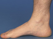
This 45-year-old male marathon runner is plagued with pain in his right heel that intermittently interferes with his intensive training programme. Pain is made worse with running, especially uphill. His ‘ankle’ feels sore and stiff in the morning but this lessens after a few minutes walking. Examination reveals a tender, fusiform swelling behind his right heel (Fig. 62.1).
1 This patient has been told that he has a tenosynovitis of the Achilles tendon. Why is this incorrect?
2 The swelling in Figure 62.1 is seen in the midportion of the Achilles tendon. How typical is this?
3 What is the role of ultrasonography in diagnosing this condition?
4 What advice would you offer this patient?
5 Discuss the conservative and operative management.
Achilles tendinopathy
Ageing and high-velocity sport predispose the tendon to injury. Typically tendinopathy occurs from overuse that places abnormal biomechanical stress on the foot and ankle. The preferred, contemporary term for tendon pain, swelling and impaired performance is tendinopathy which encompasses and supersedes the terms tendinitis and tendinosis. Traditionally it was supposed that the pain in the tendon was due to inflammation (tendinitis) but there seems to be little evidence of this and tendons which exhibit degeneration (tendinosis) are sometimes painless. Pain with this condition may be due to neovascularization irritating pain receptors.
2 Achilles tendinopathy occurs in between 7% and 9% of top-level runners. Two-thirds of recreational athletes with Achilles tendon problems have pain in the tendon (typically 2–6 cm above insertion of tendon), as seen with this man. About 23% have insertional Achilles tendon problems and 8% have pain at the myotendinous junction. In chronic Achilles tendinopathy, peritendinous swelling often develops as shown in Figure 62.1 In such cases, about 20% of the peritendinous tissue is composed of myofibroblasts which are believed to be responsible for the formation of permanent scarring around the tendon that in turn impedes circulation.
3 Ultrasonography shows discontinuity of tendon fibres and tendon thickening. Scanning also identifies neovascularization that was once thought to signify healing but paradoxically, loose granulation tissue emerges and this leads to scarring.
4 This patient should be advised that symptoms often persist for many weeks or even months. He should be advised to reduce the intensity of his activities and to warm up thoroughly in advance of running. This should be accompanied with advice to avoid walking barefoot or in flat-soled shoes such as slippers or sandals (it may be necessary to provide the patient with heel lifts).
5 In the early stages, oral non-steroidal anti-inflammatory drugs or topical gels and rest with ice and elevation are recommended. The patient should be given eccentric calf-stretching exercises. Control of biomechanical factors should be addressed with antipronatory orthoses. In recalcitrant cases, the use of dorsiflexion night splints or ‘Strasbourg socks’ should be considered although patient compliance may be an issue with these (Fig. 62.2). Because of the inherent risk of tendon rupture, steroid injections are not advocated as a routine treatment although when tendon pain does not allow eccentric stretching, a small amount of steroid, injected either side of the tendon (peritendinous), may help. Patients abstain from activity with immobilization of the ankle in a moon boot (see Fig. 56.2) or similar device.
 |
| Fig. 62.2 |
The objective of surgery is to excise fibrotic adhesions, remove degenerated nodules and make multiple longitudinal incisions in the tendon. Risks of surgery include poor wound healing and tender scars.
< div class='tao-gold-member'>
Only gold members can continue reading. Log In or Register to continue
Related posts:


Stay updated, free articles. Join our Telegram channel

Full access? Get Clinical Tree


