PROCEDURE 9 Radial Head Fractures
Open Reduction and Internal Fixation
• The fracture must be amenable to anatomic joint reduction and rigid internal fixation. This will depend on:


Indications


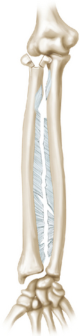
 Associated longitudinal instability of the forearm (Fig. 3)
• Fractures associated with longitudinal instability of the forearm (i.e., interosseous ligament injury)
Examination/Imaging
 Clinical evaluation
• The joint above and below the fracture should be examined for associated injury (e.g., associated distal radioulnar joint injury).
 Imaging
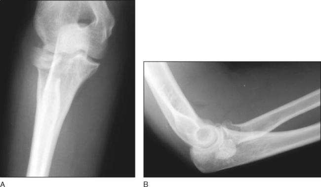
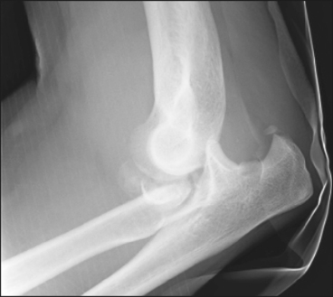
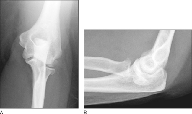
• Plain radiographs of the elbow in the anteroposterior (Fig. 4A) and lateral (Fig. 4B) planes must be obtained.
• A radiocapitellar view taken with the dorsal aspect of the supinated forearm against the x-ray plate with the beam directed 45° mediolaterally may help identify minimally displaced fractures of the radial head, coronoid, and capitellum.
• Radiographs of the ipsilateral and contralateral wrist should be obtained to determine ulnar variance if there is a question of longitudinal instability.
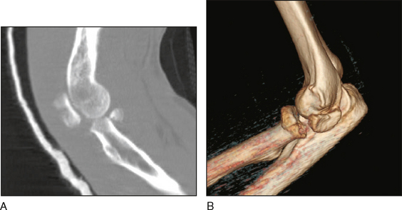
• Computed tomography scanning (Fig. 5A) with three-dimensional reformatting (Fig. 5B) allows better definition of the anatomy, the degree of comminution, and the size of the fracture fragments.
Surgical Anatomy
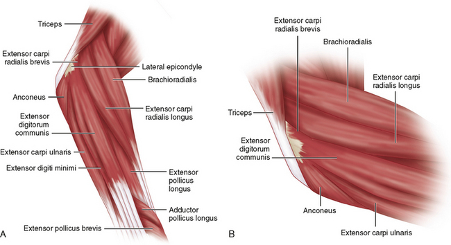
 Muscles and nerves
• Anconeus muscle/extensor carpi ulnaris (ECU) (Fig. 6A and 6B), as well as the extensor carpi radialis brevis (ECRB), extensor carpi radialis longus (ECRL), and extensor digitorum communis (EDC)
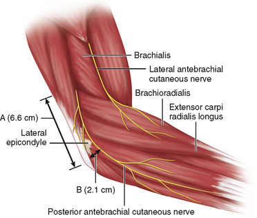
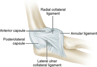
• Posterior antebrachial cutaneous nerve (PABCN; superficial and anterior to common extensor origin) (Fig. 7; LABCN = lateral antebrachial cutaneous nerve)

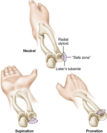
 The “safe zone”—100° arc of the circumference of the radial head that does not articulate with the radial notch of the proximal ulna (Fig. 11)