PROCEDURE 8 Terrible Triad Injuries of the Elbow

Examination/Imaging


 The elbow is inspected for obvious dislocation or open injury necessitating immediate reduction and/or surgical treatment.


 Radiographs
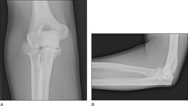
• Prereduction and postreduction (Fig. 1A and 1B) anteroposterior and lateral radiographs are obtained to examine fracture characteristics and concentricity of the elbow joint.
• Regardless of the radiographic projection, a line drawn through the center of the radial neck should intersect with the center of the capitellum.
 Computed tomography (CT)
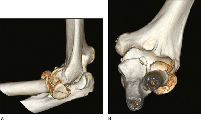
• CT is obtained following joint reduction to better evaluate fracture patterns, comminution and displacement.
Surgical Anatomy
BONES
 The proximal ulna
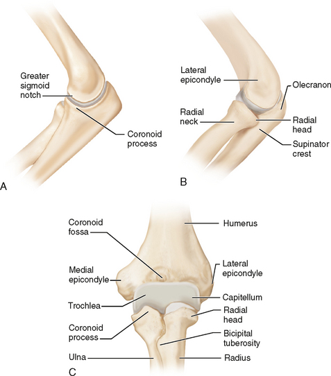
• The greater sigmoid notch, with its central guiding ridge, articulates with the trochlea and is made up of the coronoid and olecranon (Fig. 3A).
 The proximal radius
• The radial head (Fig. 3C), which is elliptical in shape and offset from the neck, articulates with the capitellum and lesser sigmoid notch.
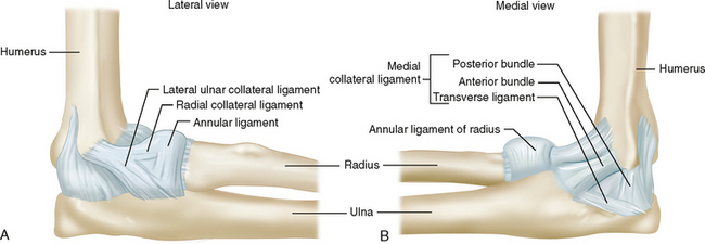
LIGAMENTS
 Lateral collateral ligament (LCL)
• The LCL consists of the lateral ulnar collateral, radial collateral, and annular ligaments (Fig. 4A).
• The lateral ulnar collateral ligament originates on the lateral epicondyle and attaches to the crista supinatoris.
• The radial collateral ligament originates on the lateral epicondyle and fans out to attach to the annular ligament.
 Medial collateral ligament (MCL)
• Its origin on the anteroinferior aspect of the medial epicondyle is distal to the axis of rotation and therefore tension increases in flexion.
• The anterior bundle originates from the medial epicondyle and inserts on the sublime tubercle of the coronoid.
• The posterior bundle originates from the medial epicondyle and inserts on the ulnar aspect of the greater sigmoid notch.
• With the arm across the chest, use a 3-L intravenous fluid bag placed under the ipsilateral scapula to protract the shoulder. A second assistant may be needed on the opposite side of the operating table.
• When using an arm table, make sure the torso is shifted close to edge of the operating table to move the elbow into the center of the table. A rolled drape placed underneath the medial or lateral aspect of the elbow with the shoulder in internal or external rotation, respectively, facilitates positioning.
• When using the lateral decubitus position, ensure the armholder is well padded and is situated over the biceps muscle to avoid pressure points. If using a bean bag for positioning leave the suction attached to maintain deflation during surgery. Tilt the operating table 10 degrees towards the operative side after the patient is secured to prevent the arm from slipping back off the arm support.
• Positioning supine with the arm across the chest and lateral decubitus positioning have an advantage over the arm table for unstable injuries as gravity tends to reduce the elbow in the former two positions, while rotation of the shoulder to gain access to the medial and lateral sides of the elbow when using an arm table tends to subluxate the elbow and stress collateral ligament repairs and fracture fixation.


Positioning
 For lateral surgery, patients can be placed in the supine position with the arm over the chest supported by a rolled drape to allow the elbow to flex to 90° (Fig. 6A).
 For medial surgery, patients can be placed in the lateral decubitus position with the aid of an elbow positioner (Fig. 6B).
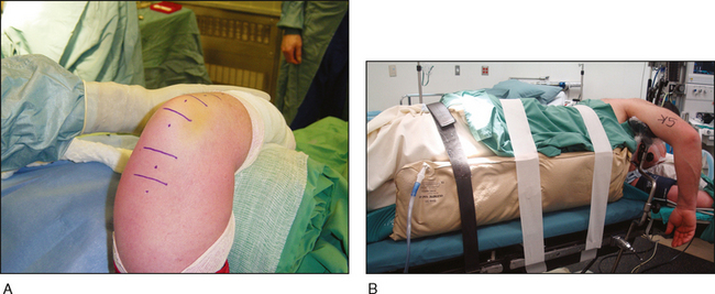
Exposures
• A sterile tourniquet will allow more proximal incision length and can be removed if more exposure is required.
• A padded armholder placed under the anterior surface of the humerus in the lateral decubitus position is needed to stabilize the arm during surgery.
SKIN INCISION
 Choice of skin incision(s) location is controversial; it depends on the fracture/instability pattern, soft tissue injury, and surgeon preference.


DEEP EXPOSURE
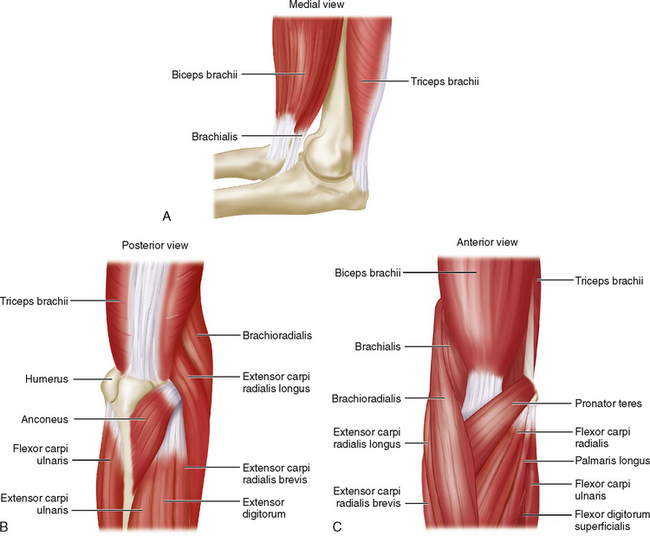
 Lateral surgical approach to the elbow: access to the radial head and neck, lateral collateral ligament, and most coronoid fractures can be achieved by one of two approaches.
• Kocher’s interval


♦ Kocher’s interval between the extensor carpi ulnaris and anconeus is identified (Fig. 7A) and incised (Fig. 7B).
Stay updated, free articles. Join our Telegram channel
Full access? Get Clinical Tree
