PROCEDURE 7 Supracondylar Humeral Fractures
Indications
 Total elbow arthroplasty (TEA) provides good to excellent results in carefully selected patients with comminuted distal humeral fractures.


Examination/Imaging
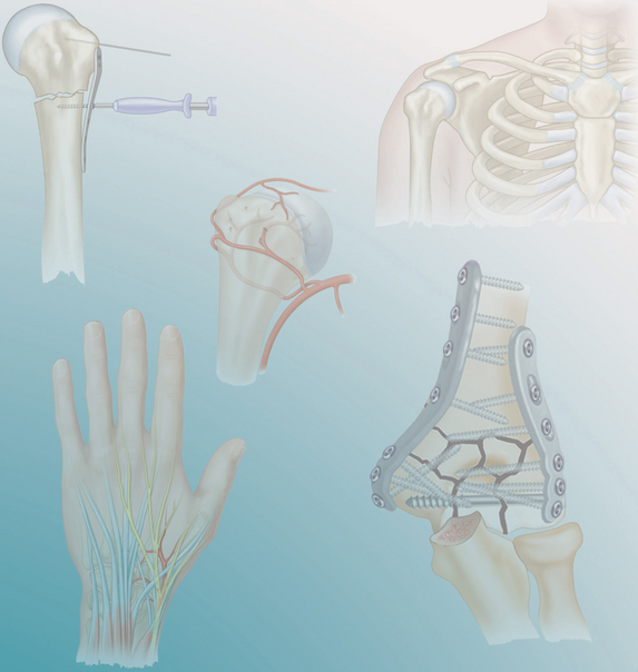
 A common pitfall is to focus immediately on the obvious injury. Examination of the shoulder and wrist is a must.


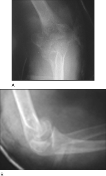
 Plain radiographs are universally the initial study of choice.
• Figure 1 shows the preoperative anteroposterior (Fig. 1A) and lateral (Fig. 1B) radiographs of a 68-year-old woman with a comminuted intra-articular distal humerus fracture.


Positioning
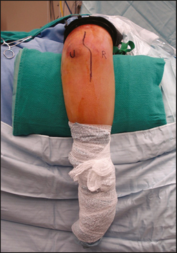
 We prefer to place the patient in the lateral decubitus position with the injured arm up (Fig. 3). The affected elbow is supported over a bolster.


Portals/Exposures
• A Hohmann retractor can be used on either side of the humeral shaft to “lift up” the shaft for exposure, rather than levering on the soft tissue excessively. This is especially important on the lateral side where the radial nerve courses proximally.

Stay updated, free articles. Join our Telegram channel

Full access? Get Clinical Tree


