PROCEDURE 6 Open Reduction and Internal Fixation of Intra-Articular Fractures of the Distal Humerus

Examination/Imaging
• Open reduction and internal fixation is the “gold standard” treatment for these intra-articular fractures.
• Total elbow arthroplasty may be considered for elderly, low-demand patients with comminuted fractures.
 Clinical examination should include: inspection of the skin for any lacerations indicating an open fracture; evaluation of median, ulnar, and radial nerve function; and examination of the wrist and shoulder for any associated injuries.

Surgical Anatomy
 Muscular anatomy (Fig. 1A): medial head, lateral head, and long head of the triceps; triceps tendon, intermuscular septum, flexor carpi ulnaris, anconeus, and extensor carpi ulnaris
 Neurologic anatomy (see Fig. 1A): radial nerve, ulnar nerve, and posterior antebrachial cutaneous nerve
 Bony anatomy (Fig. 1B): medial and lateral epicondyles, trochlea, capitellum, olecranon fossa, and olecranon
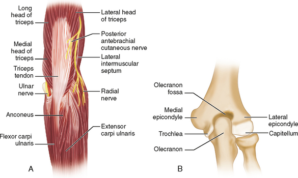
Positioning
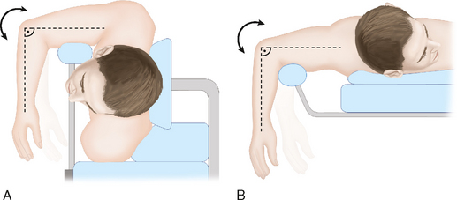
 The patient may be placed in the lateral decubitus position with the operative side facing upward (Fig. 2A) or, alternatively, the patient may be placed in the prone position (Fig. 2B).
Portals/Exposures
 Triceps-splitting approach
• A midline skin incision is made extending along the subcutaneous border of the ulna, over the olecranon and proximally in the midline of the humerus (Fig. 3A). Generous subcutaneous dissection is performed both medially and laterally to expose both epicondyles.
• The ulnar nerve is identified over the posterior aspect of the medial epicondyle (see Fig. 3A). The nerve is released both proximally and distally and retracted with a vessel loop.
• The triceps tendon and muscle are split in the midline (dotted line in Fig. 3A). The radial nerve must be identified and protected if the triceps muscle split is extended proximal to the distal third of the humerus.
• Any traumatic defects in the triceps tendon should be incorporated into the triceps split. These traumatic defects are often encountered with open fractures as the bone tears through the triceps tendon before piercing the skin.
 Triceps-sparing approach
• A midline skin incision is made similar to that used for the triceps-splitting approach and the ulnar nerve is released and retracted (see Fig. 3A).
• The ulnar nerve is followed proximally along its course over the intermuscular septum.
♦ The medial (ulnar) window is created by dissecting out the ulnar nerve and mobilizing the medial head of the triceps laterally to expose the humerus (Fig. 4A).
 Olecranon osteotomy approach
• A midline skin incision is made similar to that used for the triceps-splitting approach and the ulnar nerve is released and retracted (Fig. 5).
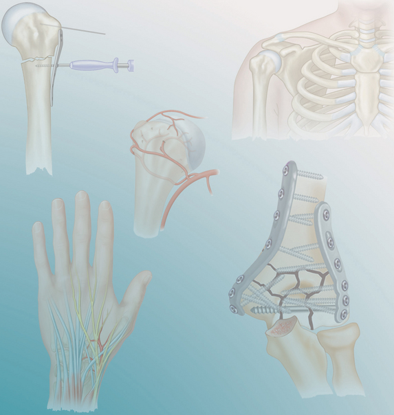
• A hole maybe predrilled through the olecranon to allow for anatomic reattachment of the olecranon at the end of the operation. This hole is made with a 3.2-mm drill bit for fixation with a 6.5-mm cancellous screw (Fig. 6A).
♦ Alternatively, two 1.5-mm Kirschner wires (K-wires) may be used to predrill holes through the olecranon and anterior cortex of the ulna, then removed prior to performing the osteotomy (Fig. 6B). This is useful if if the osteotomy is to be fixed using a tension band technique.


Stay updated, free articles. Join our Telegram channel

Full access? Get Clinical Tree


