PROCEDURE 50 Acute Total Hip Arthroplasty for Acetabular Fractures
Indications
 As a general principle, the indication for acute total hip arthroplasty (THA) is irreversible destruction of the acetabulum and/or femoral head.
 Relative indications, according to Richmond and Helfet (2003), are irreductible fracture through a single nonextensile exposure and anticipated protracted surgical time (>4 hours).
• The question that remains controversial is whether or not geriatric patients with acetabular fractures should be treated by open reduction and internal fixation (ORIF) or THA, either acute or delayed.
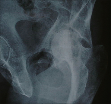
• Osteopenic patients with superomedial dome impaction—the “gull sign,” as seen in an obturator oblique radiograph of a left acetabular fracture in a 74-year-old patient (Fig. 1)—did not benefit from attempted ORIF in the Anglen et al. (2003) series.
• Nonoperative treatment maybe justified for minimally displaced fractures and specific fracture patterns: a low transverse fracture, a posterior wall fracture involving less than 25% with a stable hip, and an associated both-column fracture with “secondary congruence.”
• The author recommends ORIF of the medically fit elderly patient with an acetabular fracture in the majority of cases.
Examination/Imaging
 Relevant features of the history include evaluation for drug and alcohol abuse, comorbidities, weakness of the hip abductors, and accurate neurologic examination (sciatic nerve).
 When planning a posterior approach, blunt trauma to the gluteal muscle mass and peritrochanteric region is the most troubling finding.
 The presence of the Morel-Lavalle lesion is associated with high rates of bacterial contamination and must be addressed with surgical decompression and débridement prior to definitive fracture care.
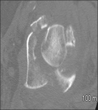
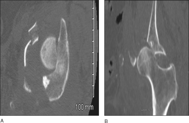
 A radiographic evaluation of the hip and pelvis includes the standard anteroposterior (AP) iliac and obturator oblique views, along with a computed tomographic (CT) scan.
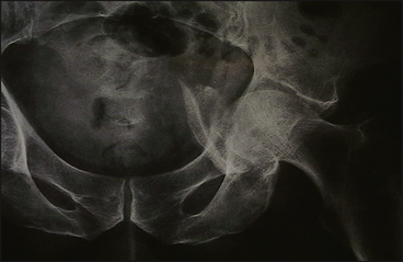
• Figure 2 shows an AP radiograph of an anterior column acetabular fracture with quadrilateral plate comminution.
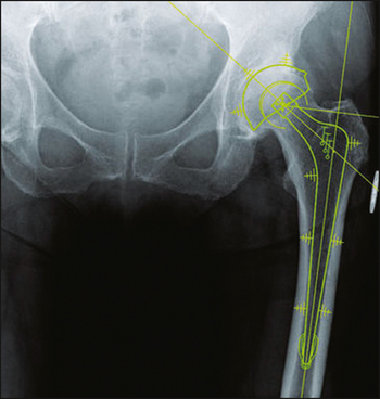
 Preoperative planning is essential; the contralateral hip should be templated for proper sizing to ensure proper implant selection and restoration of biomechanics, including offset and leg length (Fig. 5).
Surgical Anatomy
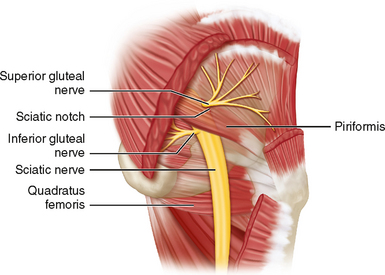
 The superior gluteal neurovascular bundle is at risk during exposure of the greater sciatic notch and sciatic buttress (Fig. 6).
• When applying traction to the gluteus medius in order to better visualize the lateral wall of the ilium, extreme caution is needed to prevent tearing of the artery or stretching of the nerve.
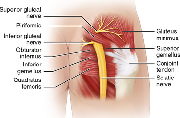
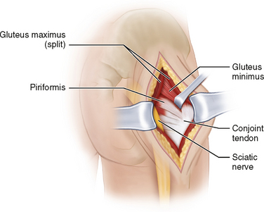
 The sciatic nerve is always at risk and must be protected at all times by positioning the knee joint in flexion and hip in extension, using special retractors, and tagging the conjoint tendon with a suture (Fig. 7).
 The medial femoral circumflex artery and its branches buried in the muscle of the quadratus femoris can be injured if this muscle is released at its insertion on the femur. However, avascular necrosis of the femoral head is not a problem when choosing THA.
Portals/Exposures
 A Kocher-Langenbeck or a posterolateral approach is recommended; it provides direct access to the posterior column/posterior wall and indirect digital access to the quadrilateral surface and pelvic brim.
 The incision begins 5 cm distal to the posterior superior iliac spine (PSIS), curves over the trochanter, and ends along the lateral aspect of the femur. The iliotibial band is incised in line with its fibers.
 The fibers of the gluteus maximus muscle are bluntly separated with two fingers, aiming towards the PSIS. Next the trochanteric bursa is incised, and the gluteus tendon may also be released for additional exposure.
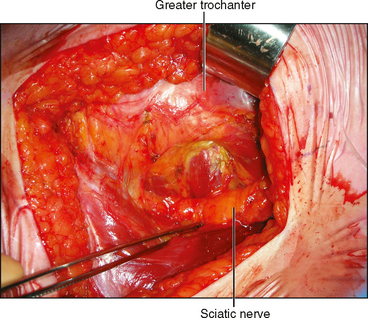
 It is imperative to identify the sciatic nerve at this point at the level of the ischion (Fig. 9), and it should be followed to its pelvic exit at the sciatic notch.
 The tendon of the piriformis muscle and the conjoined tendons (inferior/superior gemellus and obturator internus) are tagged and transected at their insertion on the trochanter (Fig. 10). The conjoined tendons will protect the sciatic nerve. The quadratus femoris muscle can also be released from its femoral insertion.