PROCEDURE 49 Fixation of Periprosthetic Femoral Fractures Using Locked Plates Combined with Minimally Invasive Insertion
• Many fractures classified as Vancouver type B1 (well-fixed stem) are in reality type B2 fractures with a loose stem that were not recognized (Lindahl et al., 2006).
Examination/Imaging
 Preoperative assessment should include range of motion of the hip and knee, soft tissue (scars) evaluation, neurovascular status, and leg lengths.
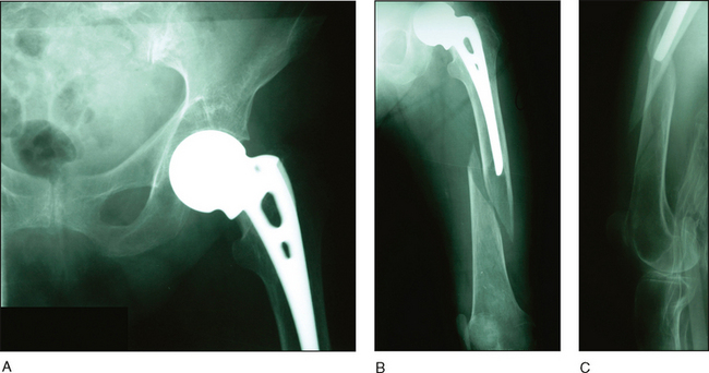
 Adequate radiographs are needed to identify the extent of the fracture, stability of the prosthesis, and the quality of the bone.
• The standard radiographs include a low anteroposterior (AP) pelvis radiograph, a frog-leg or cross-table lateral of the affected hip, and AP and lateral views of the entire femur, including the knee.
 If acetabular osteolysis is seen or suspected, then Judet films should be obtained, with a computed tomography scan to assess the extent of bony deficiency.
 Preoperative templating is necessary to determine plate length, plate contouring, and approximate screw lengths.
• The vast majority of displaced fractures are treated operatively except for the high-risk patient. Nonoperative treatment may be appropriate for stable and nondisplaced fractures. As the proximal fragment has always been a problem, many types of fixation devices have been used. Two strut allografts or a combination of one strut and one plate have proven to be the strongest constructs tested (Wilson et al., 2005).
Surgical Anatomy
 The lateral approach is a quick and easy approach that involves splitting of the vastus lateralis muscle.
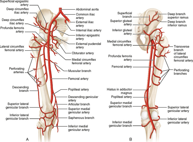
 Numerous perforating branches of the profunda femoris artery traverse the vastus lateralis muscle and can be damaged during the approach.
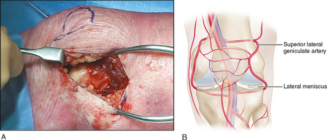
 At the level of the knee, the lateral superior genicular artery is at risk and may need to be coagulated (Fig. 3A and 3B).
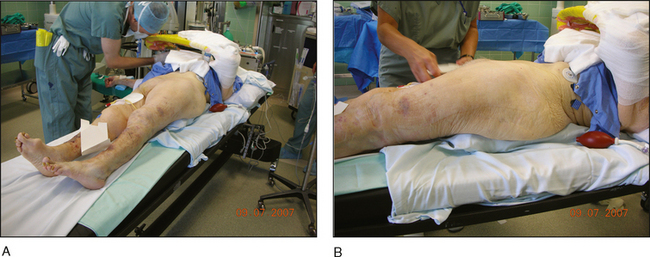
• Care must be taken during rotational alignment of the limb because the bump will cause the hip to be in an externally rotated position. An inflatable/deflatable bump can solve this problem (see Fig. 4B).
Portals/Exposures
 The surgeon begins with the small, distal, longitudinal incision (5 cm) over the lateral femoral condyle of the knee (distal window) (Fig. 5A).
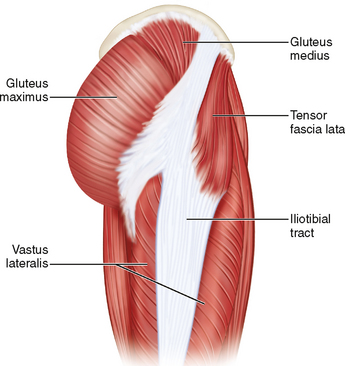
• For distal extension, it may be necessary to incise the anterior fibers of the iliotibial tract and then carry down through the capsule and synovium.
