PROCEDURE 46 Cervical Spine
Indications
 Acute fractures and dislocations that result in acute cervical spine instability require early stabilization to prevent acute or gradual displacement that can cause spinal cord compression.
• While the posterior approach offers the most predictable method of achieving a satisfactory reduction and the strongest biomechanical construct, the anterior approach allows for easier patient positioning and less extensive soft tissue dissection. Disk material and retropulsed bony fragments will require an anterior approach.
Examination/Imaging
 A detailed neurologic examination is imperative as it may influence the timing and character of the surgical treatment.
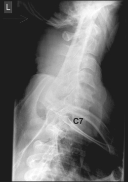
 Radiographs should be obtained in anteroposterior (AP), lateral, and swimmer’s views.
• Although computed tomography (CT) with reconstruction has replaced these standard views in some Level 1 trauma centers, they remain the standard radiologic tests for the diagnosis of significant cervical spine pathology.
 The CT scan is paramount in preoperative planning and should be used in all surgical cases. The scan should include the entire cervical spine, and continue caudally to T4.
 Magnetic resonance imaging (MRI)
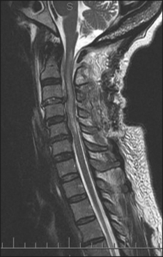
• MRI is used for evaluation of disks and ligaments. In the unconscious or sedated patient, it may be the only test that reveals significant soft tissue disruption. Additionally, the spinal cord may show evidence of injury not appreciated by other modalities (Fig. 2).
• Nonoperative treatment is acceptable in mostly osseous injuries and in the pediatric population provided that good alignment is obtained and maintained. This may range from the application of a soft collar to the installation of a halo vest. The failure rate of these modalities is higher in mostly ligamentous and soft tissue injuries. In the obese patient, bracing is less effective, and the elderly may not tolerate bracing well.
• Assure satisfactory fluoroscopic visualization of the affected levels preoperatively.
 Slightly oblique views may be necessary to visualize the lower levels if the shoulders obscure the view.
 Slightly oblique views may be necessary to visualize the lower levels if the shoulders obscure the view.
Positioning
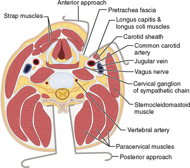
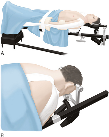
 Posterior approach (Fig. 4B)
• A Mayfield head rest, Mayfield tongs, or a halo ring is used to prevent direct pressure on the eyes during surgery. Traction is optional.
• Once the spine is reduced, traction is optional and serves to stabilize the head during the operative procedure and to counteract traction on the arms. In most cases, 5–10 lbs is satisfactory. Excessive traction on the head, fixing the spine in a distracted position, may lead to late instability and hardware failure.
