PROCEDURE 45 Open Reduction and Internal Fixation of the Acetabulum
• Nonoperative treatment may be carefully considered under certain circumstances.
 Fractures involving less than 20% of the posterior wall and with no joint incongruity on dynamic stress radiographs taken under anesthesia (Tornetta, 1999).
 Fractures not involving the posterior wall and not an associated both-column fracture pattern, evaluated out of traction or, ideally, with stress radiographs taken under anesthesia (Olson and Matta, 1993; Tornetta, 1999).
 Fractures not involving the weight-bearing articular surface as defined by roof arcs of greater than 45° on anteroposterior and both Judet views, or entering the joint greater than 1 cm below the dome condensation on computed tomography scans (Olson and Matta, 1993).
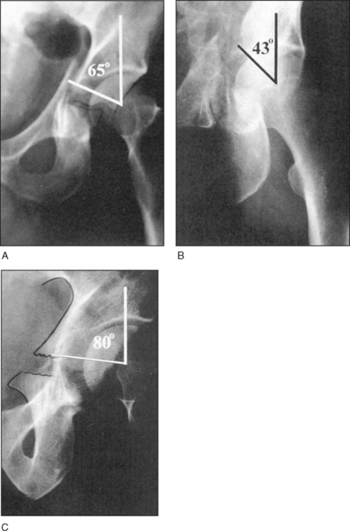
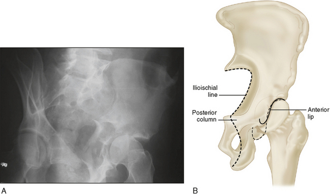
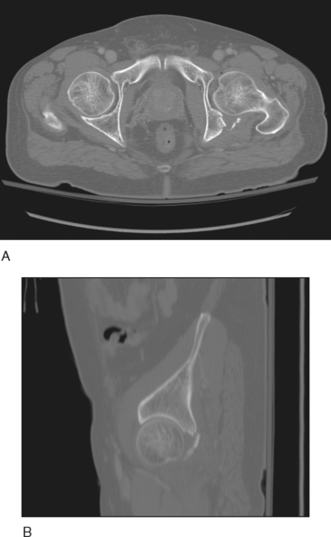
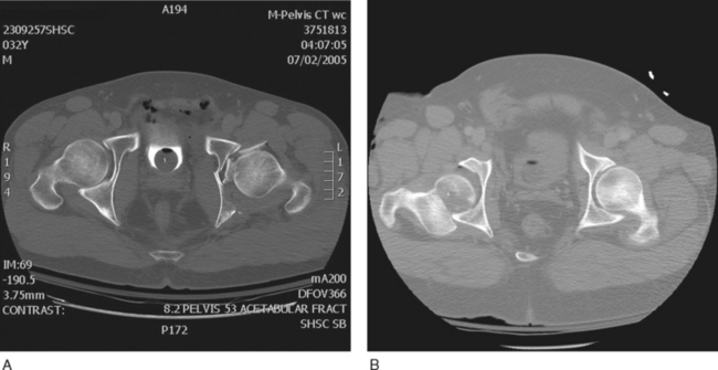
 Note: Posterior column roof arc (measured on the iliac oblique view) may need to be greater to avoid joint subluxation during weight bearing (Fig. 1A–C). Joint subluxation may occur with weight bearing if the posterior column fracture enters the joint above the ischial spine (iliac oblique roof arc of 70°), or if the anterior column fracture enters the joint above the anterior inferior iliac spine (obturator roof arc of 30°) (Vrahas et al., 1999).
 Fractures involving less than 20% of the posterior wall and with no joint incongruity on dynamic stress radiographs taken under anesthesia (Tornetta, 1999).
 Fractures not involving the posterior wall and not an associated both-column fracture pattern, evaluated out of traction or, ideally, with stress radiographs taken under anesthesia (Olson and Matta, 1993; Tornetta, 1999).
 Fractures not involving the weight-bearing articular surface as defined by roof arcs of greater than 45° on anteroposterior and both Judet views, or entering the joint greater than 1 cm below the dome condensation on computed tomography scans (Olson and Matta, 1993).
 Note: Posterior column roof arc (measured on the iliac oblique view) may need to be greater to avoid joint subluxation during weight bearing (Fig. 1A–C). Joint subluxation may occur with weight bearing if the posterior column fracture enters the joint above the ischial spine (iliac oblique roof arc of 70°), or if the anterior column fracture enters the joint above the anterior inferior iliac spine (obturator roof arc of 30°) (Vrahas et al., 1999).
Indications
 The posterior approach alone or as a component of a combined sequential or simultaneous approach may be considered in the following situations:
• Elementary and associated fracture patterns involving the posterior column (without the posterior wall)
• Operative versus nonoperative treatment for associated both-column fractures with secondary joint congruence (a fracture pattern in which the entire articular surface is free floating and no longer linked to the axial skeleton)
 Most of these injuries should be treated operatively to allow early patient mobilization and to ensure optimal articular reduction.
 “Secondary congruence” may be observed in associated both-column fracture patterns. Nonoperative treatment may be appropriate in this circumstance depending on patient factors such as advanced age and low demand. However, early mobilization may result in fracture displacement and a more difficult reconstruction at a later date.
 Most of these injuries should be treated operatively to allow early patient mobilization and to ensure optimal articular reduction.
 “Secondary congruence” may be observed in associated both-column fracture patterns. Nonoperative treatment may be appropriate in this circumstance depending on patient factors such as advanced age and low demand. However, early mobilization may result in fracture displacement and a more difficult reconstruction at a later date.
• Immediate total hip replacement
 Poor-quality bone, marked fracture impaction of the weight-bearing region, advanced patient age, and comorbidity are all considered factors that mitigate against a good outcome following fracture fixation
 Poor-quality bone, marked fracture impaction of the weight-bearing region, advanced patient age, and comorbidity are all considered factors that mitigate against a good outcome following fracture fixation
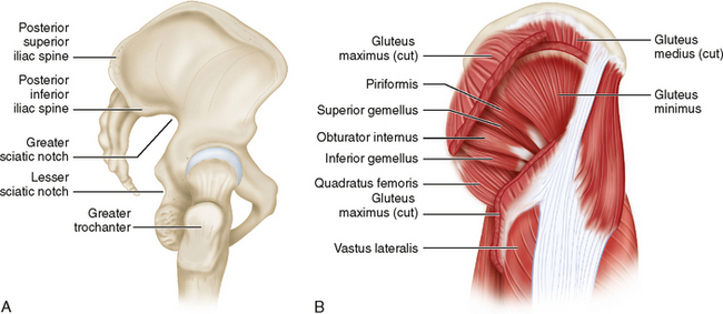
• Kocher-Langenbeck approach: may add trochanteric flip (digastrics) osteotomy to access high dome region (Siebenrock et al., 1998)
Examination/Imaging
 Preoperative planning demands a thorough understanding of all components of the fracture pattern. The surgeon must plan:
• The surgical approach (anterior, posterior, added trochanteric flip, two approaches sequentially or simultaneously, an extensile approach)
 Plain radiographs
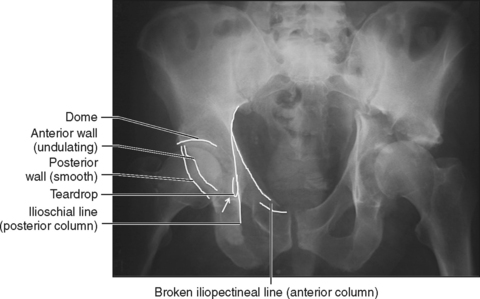
• Anteroposterior (AP) pelvis and Judet (oblique) views allow delineation of all major fracture components and are generally sufficient to plan the operative approach.
 Computed tomography (CT) scans
Surgical Anatomy
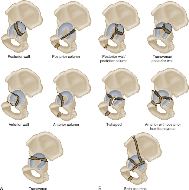
 The Letournel classification for acetabular fractures divides the most common fracture patterns into five simple types (Fig. 9A) and five associated types (Fig. 9B).
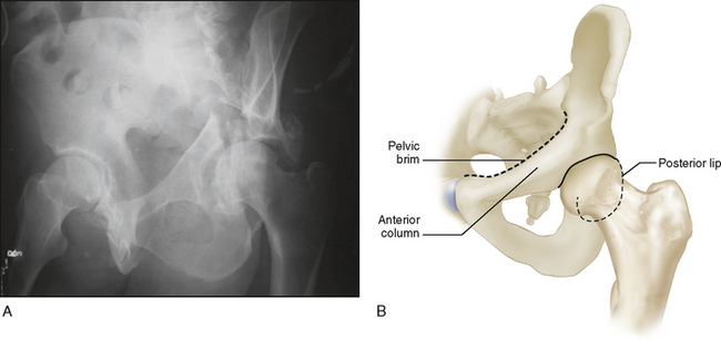
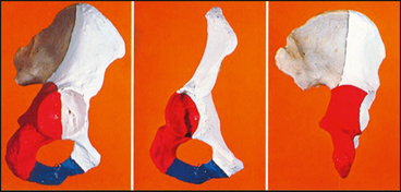
 Figure 10 illustrates the two-column concept of the acetabulum (anterior column, white; posterior column, red).